SAXS-Tensor-Tomography
Focused Xray beams can be used to scan a sample specimen with spatial resolutions from several micrometers down to a few hundred nanometers. This is the real space resolution limit one can get with such a scan, but the reciprocal space probed by the scattering in each scanning point is on the order of nanometers and below.
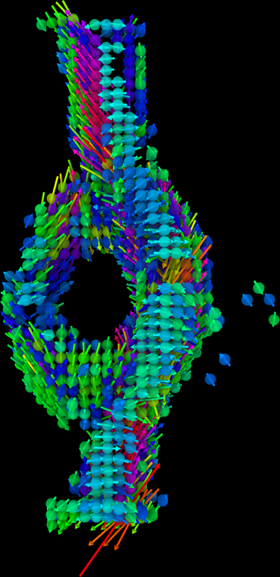
The combination of tomography, full sample scans of several dozen sample orientations, and scanning SAXS/WAXS allows the study of nanoscale features distributed over extended volumes in the specimen. Biological samples are a prime example for use of this technique as they are hierarchically structured from nano- to micro- up to centimeters. We are developing our own software solutions based on the approaches laid out by Liebi and Georgiadis et al. We sucessfully reconstructed the distribution of myelin and the 3D orientation of the nerve fibres in several thin brain slices with this technique.
M. Liebi, M. Georgiadis, J. Kohlbrecher, M. Holler, J. Raabe, I. Usov, A. Menzel, P. Schneider, O. Bunk and M. Guizar-Sicairos
Small-angle X-ray scattering tensor tomography: model of the three-dimensional reciprocal-space map, reconstruction algorithm and angular sampling requirements
Acta Cryst. (2018). A74, 12-24, https://doi.org/10.1107/S205327331701614X
Neutron sources are in their current state not yet capable of such small spatial resolution, but we were able to determine the main in-plane orientation of nerve fibres and myelin distribution in a mouse brain sample with neutrons.
Maiti, S., Frielinghaus, H., Gräßel, D. et al.
Distribution and orientation of nerve fibers and myelin assembly in a brain section retrieved by small-angle neutron scattering.
Sci Rep 11, 17306 (2021). https://doi.org/10.1038/s41598-021-92995-2