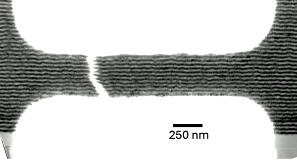
In situ tensile TEM experiments
We perform in situ tensile experiments inside the transmission electron microscope on nanometallic multilayer dogbone specimens, amongst many other possible material systems ranging from single crystals to nanocrystalline materials. Using a dedicated thermomechanical microscope holder, we applied controlled uniaxial tension, while elevating temperature and recorded real-time videos from the electron-transparent gauge section. This configuration allows us to directly observe deformation mechanisms as they unfold, in this multilayer example how cracks initiate and propagate through the layers. Because the geometry is a true dogbone, we can concentrate strain in the multilayer gauge while keeping the grips rigid, enabling direct correlation between local structural change and the imposed load path.
This capability is exciting because it unites thermomechanical loading with high-resolution imaging down to the atomic scale in one experiment. We can now answer mechanism-level questions that typically require separate tests and post-mortem inference. By quantifying the sequence and spatial location of events in the video stream, we comprehensively identify structure – mechanism – property relationships.
The straining-and-heating protocol and imaging workflow is adjustable and can apply to other material systems, such as single crystals, multiphase materials, ceramics, etc. For each, the deformation rate and thermal profile can be tuned to observe defect interactions, interface reactions, and stability limits in real time, with the option to zoom to atomic resolution to verify lattice-level mechanisms.

- Institute of Energy Materials and Devices (IMD)
- Structure and Function of Materials (IMD-1)
Room 104