Proteins in All Shapes: Research Team Reveals Diversity of Protein Conformations
Proteins in All Shapes: Research Team Reveals Diversity of Protein Conformations
12 May 2025
A single snapshot is often insufficient to understand the function of bio molecules. Proteins, for example, change their shape depending on their environment – sometimes only slightly, sometimes drastically. A research team from Forschungszentrum Jülich and Heinrich Heine University Düsseldorf has now made these changes visible by means of solid-state NMR spectroscopy. This technique allows not only individual structures to be recorded, but also entire ensembles of conformational states. The findings provide new insights into the flexibility of proteins and the dynamics of molecules that play a role in neurodegenerative diseases. The study was recently published in the Journal of the American Chemical Society.
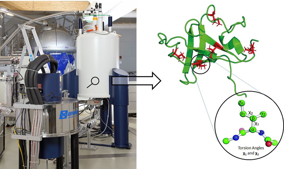
The DNP-NMR spectrometer made it possible to visualise the mobility of proteins. | Copyrights: Forschungszentrum Jülich / Limbach
More than a still image: proteins in motion
Classical structural biology aims to produce images of molecules that are as sharp and well-defined as possible, for example using X-ray crystallography. However, these methods require a molecule to be in a precise shape. In living cells, this is rarely the case. Instead, proteins move, fold, or take on transitional states – depending on the conditions.
“A static image is often insufficient. Many proteins are flexible, and to really understand them, we need to take their movements into account – in other words, the range of possible conformations they adopt,” explains Prof. Henrike Heise from Forschungszentrum Jülich’s Institute of Biological Information Processing.
Frozen diversity: what solid-state NMR reveals
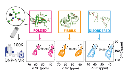
To visualize this flexibility, the Jülich team investigated proteins in frozen solution using a method that is also applied in materials research: solid-state NMR spectroscopy. The method involves shock-freezing proteins in their natural form, which has the crucial advantage of ensuring that all of the molecules' conceivable folding states remain “frozen”. In the resulting spectra, these forms leave characteristic traces that can be analysed and interpreted. The investigations were supported by state-of-the-art, high-performance equipment at the Biomolecular NMR Center, which is jointly run by Heinrich Heine University Düsseldorf and Forschungszentrum Jülich.
Specifically, the team investigated several proteins with different flexibility properties: a stably folded protein, an “intrinsically disordered” protein, which is flexible by nature, and a protein that can aggregate into amyloid fibrils in certain states – a process associated with neurodegenerative diseases.
“The analysis of the side chain of the amino acid isoleucine was particularly fascinating,” explains Leonardo Levorin, lead author of the study. “It is an excellent sensor for the dynamics and degrees of freedom at defined positions in the protein.”
By measuring the torsion angles – i.e. the rotational movements of this side chain – the team was able to show how the mobility of individual parts is related to the overall state of the protein.
New insights into protein folding
The results provide valuable insights into the dynamics of biological molecules – for example, in the folding and misfolding of proteins, which also plays a role in diseases such as Alzheimer’s and Parkinson’s. Moreover, they show how sensitive certain areas of proteins are to their environment – an interesting aspect for the development of active substances and biopharmaceuticals.