
FET.poster.png
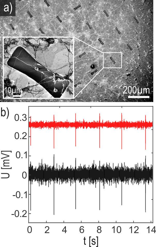
SEM images of the GFET chips after neuronal culture (a) as well as a typical recording from cardiac cells (b).
Last Modified: 17.03.2026
SEM images of the GFET chips after neuronal culture (a) as well as a typical recording from cardiac cells (b).