Pulsating Performance
Forces of Heart Muscle Cells Adapt to Changing Tissue Stiffness
Powering the blood flow through the body’s vessels is the main function of the heart. It is brought about by highly coordinated contractions of heart muscle cells – also called cardiomyocytes. Contraction rate and strength are regulated to meet the body’s demands. However, with aging or diseases the heart tissue progressively stiffens and accordingly presents increasing resistance against contractions.
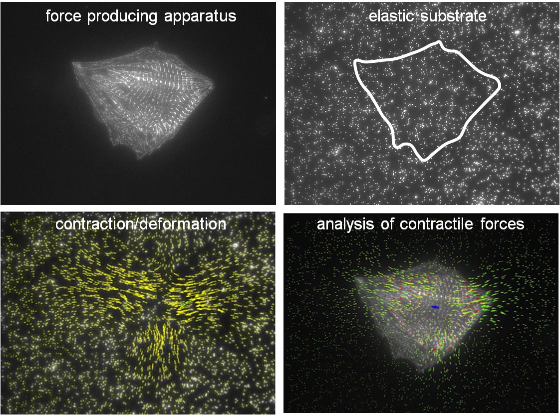
In order to mimic this situation, cardiomyocytes from rat embryos were plated on elastic substrates of various stiffness and studied with respect to morphology, structure of their force producing apparatus and total contraction force. Substrate stiffness ranged from 1 to 500 kPa thus covering elasticities of healthy and diseased heart tissue. In addition, we analyzed adhesion and shortening of the elementary contractile units, the sarcomers, within individual myofibers with high spatial resolution.
Upon substrate stiffening contraction amplitude remained almost constant. Therefore, overall contractile cell forces increase substantially with substrate stiffening. Cardiomyocytes are contraction driven and capable of adapting to changing environment, here tissue elasticity, to perform their vital function of ceaselessly pumping blood through the body.
Publication: Nils Hersch, Benjamin Wolters, Georg Dreissen, Ronald Springer, Norbert Kirchgeßner, Rudolf Merkel, und Bernd Hoffmann
The constant beat: cardiomyocytes adapt their forces by equal contraction upon environmental stiffening Biology Open 2013 BIO20133830; Advance Online Article February 1, 2013, doi:10.1242/bio.20133830

