"BrainBuilder" enables 3D reconstruction of the brain from 2D slices
“BrainBuilder” is the name of a new AI-based reconstruction method that resolves the many technical challenges involved in transforming 2D brain slices into 3D atlases. It allows for the creation of detailed, high-resolution mesoscale atlases, particularly of neurotransmitter receptors - an essential step toward better understanding the functional organization of the brain. The development of “BrainBuilder” involved an international team of researchers. Their study has now been published in the journal Communications Biology.
Mesoscale brain structures include cortical layers and their building blocks, known as cortical columns. These structures can be visualized at high resolution with post-mortem 2D imaging, for example through histological staining or receptor autoradiography. Of particular interest is the distribution of neurotransmitter receptors, which play a central role in information processing and signal transmission, and which can be visualized using receptor autoradiography. Mesoscale atlases play a crucial role in elucidating the brain’s complex architecture, providing insights into fine structural details essential for advancing our understanding of both normal brain function and pathological changes.
Until now, 3D reconstruction of receptor autoradiographies from 2D slices—so-called “sections”—has been impossible. Simply “stacking” the slices usually does not yield a correct 3D image, since the sections may be cut at varying angles and intervals, may be missing, deformed, or acquired with different methods. The research teams from Jülich, Düsseldorf, London, Montreal, and New York therefore set out to develop an (automated) technique for precise 3D reconstruction.
The result is the pipeline presented in the study: “BrainBuilder”—a software tool that combines various steps of image processing and analysis, aligning them in such a way that a usable brain model is created. AI plays an important role here: at its core is a deep learning network (U-Net) trained on synthetic data. “BrainBuilder” is highly flexible: it can handle different data types, missing sections, different tissue types, and even work without a matching reference brain image—and it is applicable across species.
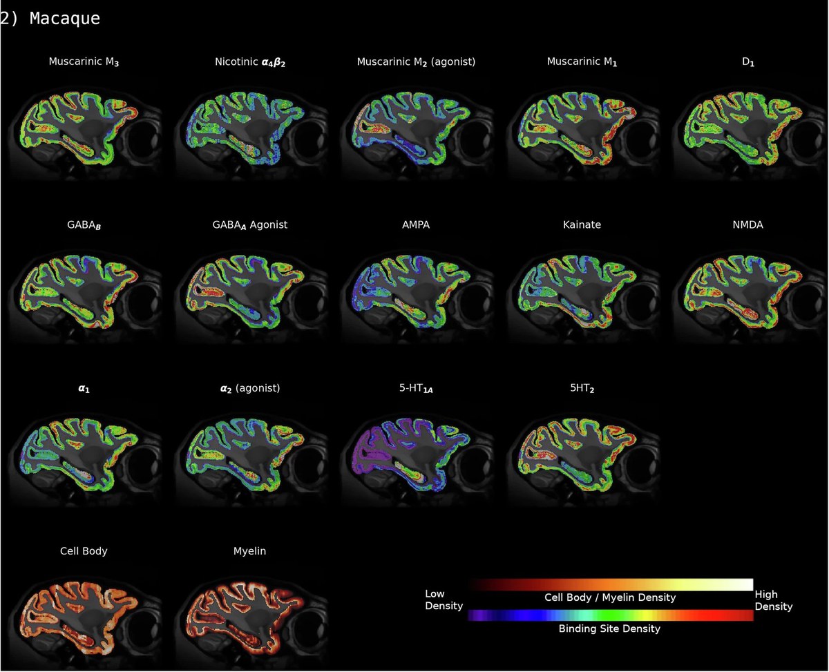
To validate the approach, the team tested the pipeline on two large datasets of brain slices: one covering the entire human brain, and one from the brain of a macaque. These datasets, collected over more than twenty years by neuroscientists Karl Zilles and Nicola Palomero-Gallagher at Heinrich Heine University Düsseldorf and Forschungszentrum Jülich, show in detail the distribution of various receptors for “classic” neurotransmitters such as glutamate, GABA, acetylcholine, noradrenaline, serotonin, dopamine, and adenosine.
Using “BrainBuilder,” the researchers successfully reconstructed the volume and distribution of 20 receptors in one hemisphere of the human brain; in the macaque brain, they reconstructed a selection of 14 receptors, along with cell bodies and myelin-stained sections. An MRI scan was available for the human brain as a reference, but not for the macaque brain.
One key outcome highlighted by the scientists is that “BrainBuilder” makes it possible to reconstruct a wide range of 2D brain datasets at the mesoscale level in 3D. This opens new opportunities in neuroscience to image the brain in even greater detail in specific regions, enabling cross-species comparisons and a deeper understanding.
Original publication:
Funck, T., Wagstyl, K., Lepage, C. Palomero-Gallagher, N. et al. Brainbuilder: a software pipeline for 3D reconstruction of cortical maps from multi-modal 2D data sets. Commun Biol 8, 1015 (2025). https://doi.org/10.1038/s42003-025-08267-6
Contact

apl-.Prof. Dr. rer. nat. Nicola Palomero-Gallagher
Working Group Leader "Receptors"
- Institute of Neurosciences and Medicine (INM)
- Structural and Functional Organisation of the Brain (INM-1)
Room 2005b
Press Contact

Erhard Zeiss
Wissenschaftlicher Kommunikationsreferent
- Institute of Neurosciences and Medicine (INM)
- Structural and Functional Organisation of the Brain (INM-1)
Room 3033