Premotor Cortex Remapped: Seven Subareas and Functional Distinction
Researchers from the Institute of Neuroscience and Medicine (INM-1) and the Cécile and Oskar Vogt Institute for Brain Research have remapped the human premotor cortex, identifying seven clearly distinguishable subareas. The new histologically high-resolution maps show how the different regions are anatomically delineated. This new subdivision helps clarify the functional differences between these regions. The new maps are available in the Julich Brain Atlas, a core component of EBRAINS—the European digital research platform for neuroscience. The study has now been published in Communications Biology.
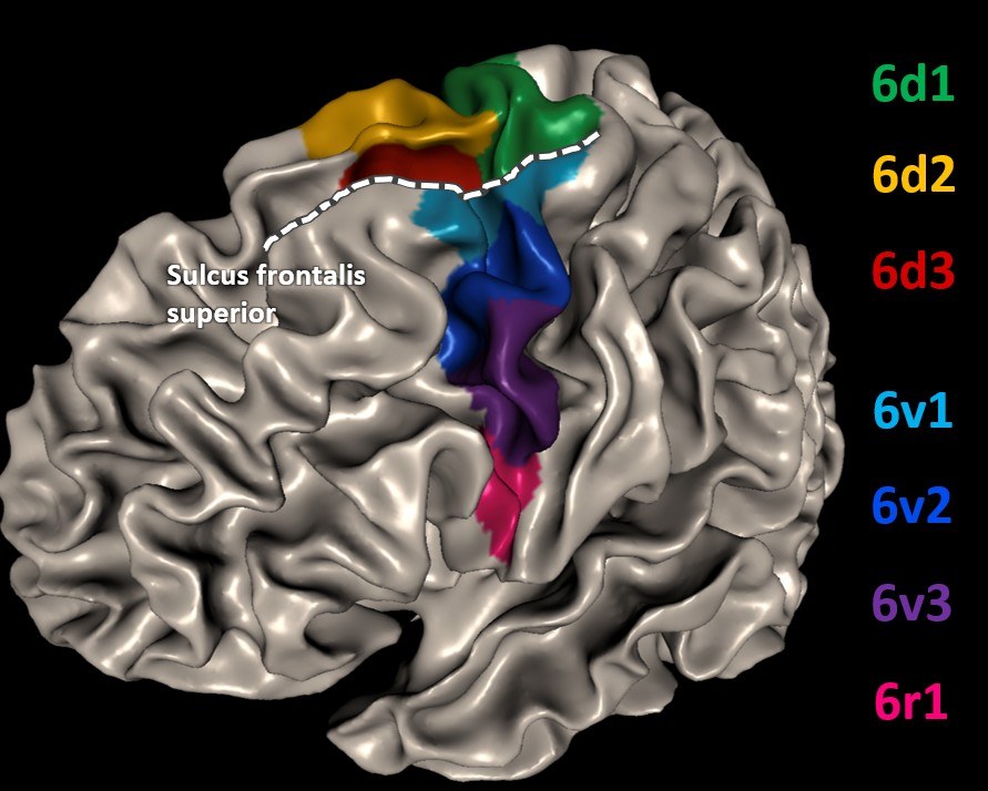
The premotor cortex is a region of the cerebral cortex that prepares movements by linking sensory information to the corresponding motor actions—for example, when one sees a cup and then reaches for it. The current study demonstrates that there is now also an anatomical correlate to the functional specialization of the premotor cortex: a dorsal (upper) and a ventral (lower) region, each consisting of several brain areas. These anatomically distinct regions can be easily identified by a brain sulcus in the frontal lobe called the superior frontal sulcus (Sulcus frontalis superior). The findings show that leg and arm movements are predominantly associated with the dorsal areas, whereas grasping and mouth movements are primarily located in the ventral regions.
Another key finding of the study is that the researchers were able to determine that the central frontal eye fields (FEF), responsible for eye movements, belong to the premotor cortex—and not, as often assumed, to the anterior prefrontal cortex.
The new premotor maps are based on histologically high-resolution data from ten human brains and provide a precise anatomical foundation for better interpretation of imaging data from healthy subjects or patients. They thus form a basis for medical applications such as planning neurosurgical interventions for epilepsy or brain tumors, helping preserve essential motor and cognitive functions.
Original publication:
Ruland, S.H., Sigl, B., Stangier, J., Caspers, S., Bludau, S., Mohlberg, H., Pieperhoff, P., Amunts, K. Revised cytoarchitectonic mapping of the human premotor cortex identifies seven areas and refines the localisation of frontal eye fields. Commun Biol 8, 1143 (2025). https://doi.org/10.1038/s42003-025-08528-4
Contact

- Institute of Neurosciences and Medicine (INM)
- Structural and Functional Organisation of the Brain (INM-1)
Room R 3034
Press Contact

Erhard Zeiss
Wissenschaftlicher Kommunikationsreferent
- Institute of Neurosciences and Medicine (INM)
- Structural and Functional Organisation of the Brain (INM-1)
Room 3033