Autoradiography
In vitro techniques
- Autoradiography
- Immunohistochemistry
- Mass spectrometry
Many physiological mechanisms as well as numerous neurological diseases involve the complex interaction of various receptors. Quantitative evaluation of the receptor distribution is essential for developing improved procedures in diagnosis and effective treatments of neurological diseases.
Quantitative Receptor Autoradiography
Using in-vitro autoradiography we analyze a broad range of different neuroreceptors in brain slices (10 -20 µm thick). After preincubation in buffer to remove endogenous ligand, sections are incubated with a radioactive substance, that binds specificly to the receptor being studied (radioligand). For the assessment of non-specific binding, selected slices are incubated with the radioligand in presence of an specific antagonist. After washing and a rapid rinse in ice-cold water the sections are dried and exposed against phosphor-imaging plates or conventional film slides in combination with tritium activity standards (see figure 1) for quantitative evaluation. Specific binding is calculated as the difference of total and nonspecific binding. Adjacent sections are stained for cytoarchitectonic identification of brain nuclei / cells. Figure 2 shows the binding of a radioligand specific for A1 adenosine receptors next to the histological staining of the same brain region.
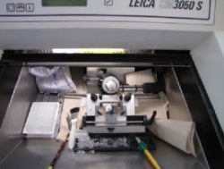
Fig. 1:
(A) Cutting of brain tissue using a cryostat.
(B) Autoradiographic procedure (from left to right): preincubation of slices, incubation with radioligand (main incubation) and 3 washing steps at 4°C.


Fig. 2: Definition of regions in autoradiography: via histological stainings (right) it is possible to quantify the receptor density in regions of interest (left. Autoradiogram with [3H]DPCPX as radioligand specific for adenosinergic A1 receptors).
In-vitro binding experiments have the advantage that the blood-brain barrier can be avoided and metabolic processes are reduced. Therefore, kinetic parameters can be reliably determined and be used for the characerization of new potential PET ligands. We perform competition and saturation studies on brain slices as well as on brain homogenates: pharmacokinetic parameters like the binding potential (Bmax) and the dissociation constant (KD) are defined and selectivity of the radioligand is tested by applying selective competitors. Examples for saturation
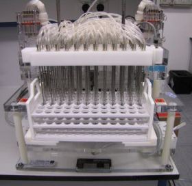
Fig. 3:
Cell harvester for saturation and competition studies on brain homogenates.
(A) Saturation analysis with scatchard plot of [3H]CPFPX for the evaluation of Bmax and KD.
(B) Competition analysis with adenosinergic A1 receptor antagonists (CPFPX, DPCPX, N-0840) und A2A receptor antagonists (ZM 241385).

Immunhistochemistry
Spatial resolution archieved in brain slices of autoradiographic experiments is higher than in in-vivo imaging techniques like PET. Still, the differentiation between various cell types is only possible by using immunohistochemical methods with specific antibodies. In figure 4 the binding of [3H]CPFPX, a specific adenosinergic A1 receptor ligand, in a rat brain slice is shown. In addition different immunohistochemical stained cell types are shown for comparison.


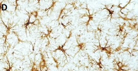
Fig. 4: (A) Region-specific binding of [3H]CPFPX to adenosinergic A1 receptors. Cell-specific binding via immunohistochemistry: Neuronal nuclei as marker for neurons (B), adenosinergic A1 receptors (C) and glial fibrillary acidic protein as marker for glia cells (D).
Laser ablation mass spectrometry with inductive coupled plasma ion source (LA-ICP-MS)
For the quantitative analysis of elements we apply LA-ICP-MS in brain tissue of humans and different animal models. This technique was developed in the Brainmet lab (Forschungszentrum Jülich) for a wide range of biomedical applications. The cooperation is based on the quantification and systematic image analysis as well as on neuro-scientific questions (see figure 5).

Fig. 5: Example of LA-ICP-MS for the validation of an animal model for parkinson’s disease. The cerebral distribution of iron (Fe), copper (Cu) and zinc (Zn) was determined in a mouse, whose substantia nigra was lesioned unilaterally via the injection of 6-OHDA.

Fig. 6: Drug monitoring of the platinum concentration in a hair sample of a tumor patient controlled by medication with cisplatin (4 cycles).