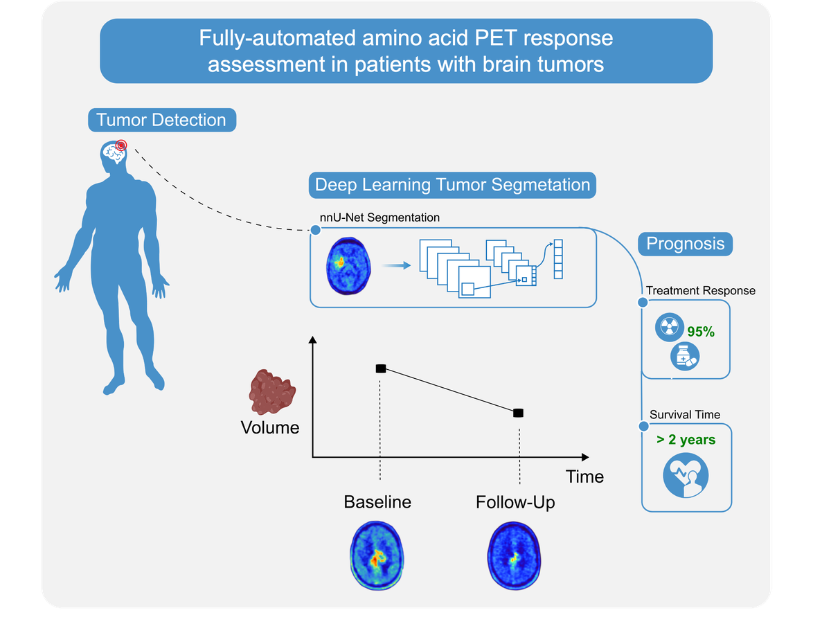
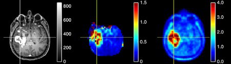
BRAIN TUMOURS
About
The brain tumour group focuses on the preclinical and clinical evaluation of new radiopharmaceuticals for nuclear medicine diagnostics of patients with brain tumours (e.g., gliomas and metastases) using positron emission tomography (PET) and the correlation and investigation of advanced magnetic resonance imaging (MRI) techniques. This includes the assessment of amino acid uptake in brain tumours, along with modern developments in functional MRI, including structural and functional connectivity and the clinical application of correlative hybrid PET/MRI for diagnosis, therapy planning and therapy response assessment in brain tumour patients.
Further, to enhance the potential of PET/MRI for brain tumour diagnostics, quantitative image analysis methods, including artificial intelligence technologies such as radiomics and deep learning, are being developed and evaluated.
Additional fields of specialist interest include the evaluation of the therapeutic effects of novel amyloid-β binding peptide ligands for neurodegenerative diseases such as Alzheimer’s using behavioural tests, autoradiography, as well as PET and MRI.
Research Topics
- Amino acid PET
- Advanced MRI
- Hybrid PET/MRI
- Quantitative image analysis & artificial intelligence
- Functional connectivity in brain tumour patients
- Preclinical brain tumour models
- Neurodegenerative diseases
HIGHLIGHTS
AI-based tumour detection and segmentation for an automated assessment of therapy response
A new artificial intelligence tool provides a fully automated, easy-to-use and objective way to detect and evaluate brain tumors. Developed for use with amino acid PET scans, the deep learning-based segmentation algorithm can assess brain tumor patients’ response to treatment with quality comparable to that of an experienced physician, but in a fraction of the time.
Correlative spectroscopic MR imaging and FET PET in brain tumours using hybrid PET-MRI
Both FET PET and spectroscopic MR imaging (MRSI) can identify brain tumor tissue with higher accuracy than conventional MRI. In a hybrid PET-MRI study on 30 untreated patients with various brain tumors, FET enhancement was compared with the MR spectroscopic result. The analysis was carried out at the anatomical coordinates at which control results from stereotactic biopsies were determined as part of the study. There was a slightly higher accuracy of FET uptake compared to the choline/N-acetyl aspartate ratio.