Dedicated diffusion phantoms for the investigation of free water elimination and mapping: insights into the influence of T2 relaxation properties
2nd March 2020
Ezequiel Farrher, Farida Grinberg, Li-Wei Kuo, Kuan-Hung Cho, Richard P. Buschbeck, Ming-Jye Chen, Husan-Han Chiang, Chang-Hoon Choi, N. Jon Shah
In diffusion-weighted magnetic resonance imaging (DWI), specific MRI sequences are used to measure the random motion of water molecules within a voxel of tissue. Due to the sensitivity of molecular displacement to the surrounding environment, DWI can be used to characterise details relating to tissue architecture at a microscopic level and is consequently useful in both fundamental research and clinical applications relating to stroke, tumours and various other neurological disorders.
Diffusion tensor imaging (DTI) is a method developed to analyse DWI data that provides rotationally invariant parameters that can be used to infer different properties of the tissue microstructure. This enables, among several other things, a map of the axonal network in the brain to be created and allows the identification of areas of disruption indicative of pathology.
However, DTI metrics can only be considered to be tissue specific if the voxel under analysis contains a single type of tissue. The aim of this work is to introduce the design, characterise the NMR properties and demonstrate the use of two dedicated anisotropic diffusion fibre phantoms, useful for the study of free water elimination (FWE) and mapping models. In particular, the recently proposed FWE DTI approach with explicit account for differences in the transverse relaxation times between the free water and tissue compartments is investigated.
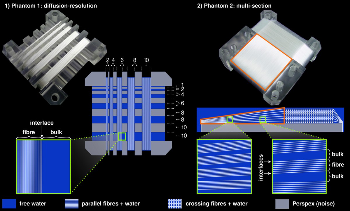
The figure above shows photographs and schemes of the phantoms. A, phantom 1. Diffusion-resolution phantom with five sections of bundles of parallel fibres of increasing thickness from left to right. The sections of fibres are separated by alternating compartments of free water and Perspex of increasing sizes. The numbers in the picture indicate the dimensions in millimetres. B, phantom 2. Multi-section phantom containing four sections with different fibre configurations. The section of interest for this work (red ROI) consists of nearly parallel fibre layers, which go from an area of high packing density (left-hand side) to an area of low packing density (right-hand side).
Original publication: