Effect of Zolpidem in the Aftermath of Traumatic Brain Injury: An MEG Study
24th March 2020
Praveen Sripad, Jessica Rosenberg, Frank Boers, Christian P. Filss, Norbert Galldiks, Karl-Josef Langen, Ralf Clauss, N. Jon Shah
Zolpidem is a drug commonly used to treat insomnia, but it has also been shown to have a paradoxical therapeutic affect in various disorders of consciousness, such as traumatic brain injury, dystonia and Parkinson's disease. Consequently, there is great interest in how zolpidem affects the brain when administered to patients affected by neurological disorders.
In this case study, magnetoencephalography (MEG) is used to investigate a fully conscious, ex-coma patient who suffered from neurological difficulties due to traumatic brain injury. Directly following the injury, the patient took zolpidem for several years and his symptoms relating to speech and motor function were seen to improve dramatically. Here, in order to further understand this rather counterintuitive effect of zolpidem, differences in the spectral power and MEG functional connectivity at the source level are measured using a weighted phase lag index (WPLI) in the resting state brain recordings of the patient before and after intake of zolpidem.
The findings of the study show a reduction in MEG power in the theta-alpha band (4–12 Hz) and an increase in the frequency band (20–43 Hz) following zolpidem intake. Changes in the cortico-cortical functional connectivity after zolpidem intake were also observed. These findings support the assumption that zolpidem has an effect on neuronal activity and consequently highlights the need for larger cohort studies in order ascertain the future role of zolpidem in the treatment of patients affected by neurological disorders. Furthermore, the study highlights the efficacy of resting state MEG as an investigative tool for traumatic brain injury.

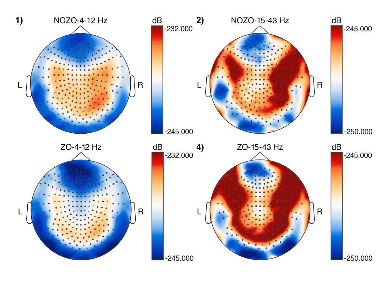
The figure above shows the power spectral densities across the sensor topography for 4–12 Hz and 15–43 Hz frequency bands for the patient with (ZO) and without Zolpidem (NOZO). Differences in PSD values in the left frontal areas (neurological orientation) before and after ZO under frequency 15–43 Hz can be seen.
Original publication:
Effect of Zolpidem in the Aftermath of Traumatic Brain Injury: An MEG StudyOriginal publication:
