High-resolution and low dose characterization

Quantitative high-resolution transmission electron microscopy

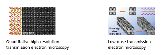
Transmission electron microscope utilizes fast electrons as illumination source to “see” atomic information (e.g., atom positions, atom varieties, valence, etc) in a material. Like other imaging systems, e.g., human eyes, light microscopes, electron microscope suffers from imaging imperfections as well, largely resulted from lens aberrations. Although hardware aberration correctors developed recently allow scientists to extend the point resolution of an electron microscope with field emission gun to its information limit, true material information can only be obtained, if an experimentally recorded image can be reproduced quantitatively by image simulations.

For quantitative high-resolution transmission electron microscopy, we mainly use a technique called negative spherical aberration imaging (NCSI), invented by our emeritus colleague, Dr. Chunlin Jia, and co-workers. A detailed description can be found elsewhere.

The image quantification relies on the absolute image contrast matching between experimental and simulated images. By doing so, the intrinsic information of materials can be extracted from those unwanted (so-called artefacts) induced by imaging imperfections (such as sample mistilt and residual lens aberrations). See also here.

Low dose transmission electron microscopy

Transmission electron microscopy (TEM) is an essential tool for characterizing the nanoscale and atomic structures of materials, offering critical insight into their fundamental physical properties. Modern TEM has been revolutionized by the development of hardware spherical and chromatic aberration correctors, which, in combination with high-brightness electron sources, monochromators and excellent mechanical and thermal stability, allows deep-sub-Ångstrom spatial resolution to be achieved with high beam current density, bringing new opportunities for tackling topical problems in materials science. This progress inevitably results in new challenges for studies of beam-sensitive materials, which require a low electron irradiation dose to avoid beam-induced structural alteration, thereby limiting the achievable signal-to-noise ratio.

Beam-sensitive materials, which often contain low-Z elements, include but are not limited to organic crystals, polymers, hybrid organic-inorganic (OI) materials and even some inorganic materials such as hydroxides. They comprise systems that are of interest in a broad range of scientific and engineering disciplines, including energy, pharmaceutical and environmental sciences.

We are currently developing low dose techniques, for example, integrated differential phase contrast imaging and electron ptychographic diffractive imaging. Knowledge learnt from life science also inspired us to carry out studies on cryogenic operation, automation, and fast detection.

Research Topics:

Quantitative high-resolution transmission electron microscopy, low dose transmission electron microscopy, structure-property relationship

High-resolution and low dose characterization

Points of contact

Dr. Lei Jin

Scientific staff at ER-C-1

  • er-c
  • er-c-1
Building 05.2 /
Room 3082B
+49 2461/61-2413
E-Mail

Penghan Lu

Scientific Staff at ER-C-1

  • er-c
  • er-c-1
Building 05.7 /
Room 2016
+49 2461/61-85121
E-Mail

Related publications

Publications

K. Ran, F. Zeng, L. Jin, S. Baumann, W.A. Meulenberg, and J. Mayer. “in situ observation of reversible phase transitions in Gd-doped ceria driven by electron beam irradiation”. Nature Commun. 15, 8156 (2024). Link: https://www.nature.com/articles/s41467-024-52386-3

K.W. Urban, J. Barthel, L. Houben, C.L. Jia, L. Jin, M. Lentzen, S.B. Mi, A. Thust, and K. Tillmann. “Progress in atomic-resolution aberration corrected conventional transmission electron microscopy (CTEM)”. Prog. Mater. Sci. 133, 101037 (2023). Link: https://www.sciencedirect.com/science/article/pii/S0079642522001189

L. Jin, F. Zhang, F. Gunkel, X.K. Wei, Y.X. Zhang, D.W. Wang, J. Barthel, R.E. Dunin-Borkowski, and C.L. Jia. “Understanding structural incorporation of oxygen vacancies in perovskite cobaltite films for electrocatalysis”. Chem. Mater. 34, 10373-10381 (2022). Link: https://pubs.acs.org/doi/10.1021/acs.chemmater.2c02043

Z.H. Ge, W.J. Li, J. Feng, F. Zheng, C.L. Jia, D. Wu, and L. Jin. “Atomic-scale observation of off-centering rattlers in filled skutterudites”. Adv. Energy Mater. 12, 2103770 (2022). Link: https://onlinelibrary.wiley.com/doi/full/10.1002/aenm.202103770

S. You, P.-H. Lu, T. Schachinger, A. Kovács, R.E. Dunin-Borkowski, and A.M. Maiden. “Lorentz near-field electron ptychography”. Appl. Phys. Lett. 123, 192406 (2023). Link: https://pubs.aip.org/aip/apl/article/123/19/192406/2920259

F. Allars, P.-H. Lu, M. Kruth, R.E. Dunin-Borkowski, J.M. Rodenburg, and A.M. Maiden. “Efficient large field of view electron phase imaging using near-field electron ptychography with diffuser”. Ultramicroscopy 231, 113257 (2021). Link: https://www.sciencedirect.com/science/article/pii/S0304399121000498

Last Modified: 07.03.2025