Advanced Scanning Probe Microscopy
We are a diverse team investigating energy materials and electrocatalysts by means of advanced Atomic Force Microscopy (AFM) techniques and nanoindentation. Force-distance-curve and current-sensitive AFM modes reveal nanomechanical and nanoelectrical properties in electrolyser and battery materials. Electrochemical Strain Microscopy (ESM) explores Li-ion mobility through electro-chemo-mechanical coupling in battery components like cathodes, anodes, and solid electrolytes. These techniques are complemented by nanoindentation analysing mechanical properties. Thereby, we unravel correlations between structural, electrical, chemical, and mechanical properties. Additional in-operando analysis facilitated by the use of home-built electrochemical cells sheds light into time and electrochemical load dependent phenomena, such as the solid-electrolyte-interphase evolution in lithium battery anodes.
Atomic Force Microscopy (AFM) and Nanoindentation
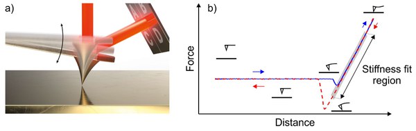
Atomic Force Microscopy is a versatile technique to be applied in various environments, from Ultra-high vacuum to electrochemical cells. The deflection of a cantilever with a laser aligned on its backside above the tip is detected by a split photodiode, which translates the motion of the cantilever into a voltage signal. A piezo electric scanner moves the cantilever based on the sensed voltage signals, allowing a sub-nanometer precise control. In dynamic modes the cantilever oscillates, e.g. in the PeakForce Tapping Mode (Bruker, USA). The cantilever is approached and retracted towards and from the sample. During this cycle, attractive or repulsive forces are imposed onto the cantilever. The signal can then be converted to force-distance curves and nanomechanical sample properties, such as stiffness, are calculated. Simultaneously, utilizing conductive tips and additional current-sensing modules, the nanoelectrical properties of samples are assessed.
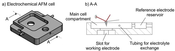
Besides ex-situ analysis, AFM measurements are performed in-situ/in-operando. We utilize home-built electrochemical AFM cells for single crystals and thin films. The insertion of the working electrode seals the cell and allows to fill the cell compartments with electrolyte. The electrolyte can be exchanged during experiments via tubings and a separate reservoir for the reference electrode reduces possible contamination and ensures a stable reference potential.
Another advanced AFM technique that we use in our lab is the Electrochemical Strain Microscopy (ESM). This mode is based on electro-chemo-mechanical coupling providing information about the local ionic conductivity. It is employed foremost in studying battery materials, from cathodes, and anodes to solid-state electrolytes. 1 We contribute to a better understanding of the complex signal origin process and explore applications of ESM especially correlatively with other techniques such as near edge X-ray absorption fine structure (NEXAFS) and nanoindentation. 2
While AFM usually probes the topmost sample surface with displacements of a few nm, instrumented nanoindentation indents the sample with displacements up to a few µm. Overall, our group covers a broad range of high-resolution characterization techniques. In the following, example studies on electrolyzer and battery energy materials are presented.
Electrolyser
Green hydrogen, produced using renewable energy sources, is fundamental for the transition to sustainable energy systems. While water electrolysis, the process of producing hydrogen from water using electricity, is a well-established technology, the high manufacturing and production costs prevent its commercial breakthrough. Our research and development efforts focus on understanding aging phenomena of these systems and enhancing durability.
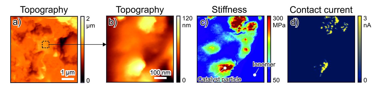
In an AFM and nanoindentation study on a long-term operated electrolyzer core material, more precisely the proton exchange membrane electrode assembly anode, force-distance-curve based and current-sensitive AFM analysis analyzed the anode aging. 3 AFM revealed the nanoscale distribution of catalyst and ionomer surface species. The stiffness map reveals stiffer domains indicating the presence of catalyst particles which are electrically conductive as indicated in the respective contact current map.
In order to boost the performance of metal-air battery systems and all-solid-state batteries towards high energy densities, longer lifetime, and improved cycling stability at the same time as thermal stability and safety, an exact understanding of mechanisms and processes in battery materials under electrochemical load is necessary. Degradation, interfacial properties and intercalation/deintercalation occur on a molecular level and hence, high-resolution microscopy is employed by advanced AFM, correlatively with other techniques. To characterize air and moisture sensitive battery materials as well, such experiments are conducted within a glove box system.
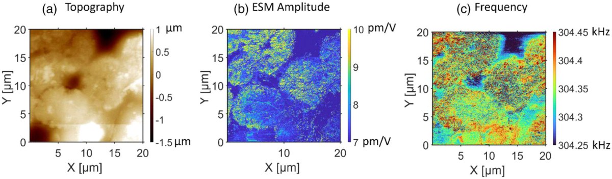
In one of our studies on the Li-ion battery cathode material NCM622 (LiNi0.6Co0.2Mn0.2O2), ESM was employed to understand the Li-ion mobility, and nanoindentation to relate the mechanical properties exhibited by the material to the chemical state and ion mobility 4. The topography exhibits an ensemble of NCM622 particles which are at 0% state of charge (SOC). The simultaneously recorded ESM amplitude map reveals increased Li-ion mobility within the particles compared to the binder. Moreover, amplitude variations are observed within the individual particles.
Contact

Last Modified: 04.04.2026