SEQUENCE & SCIENTIFIC COMPUTING
The sequence development team focuses on the design of new magnetic resonance imaging (MRI) techniques tailored to neuroscientific applications. MRI sequences drive the magnetic fields to manipulate the spin system with the aim of generating high contrast images of tissue in short scan times.
However, the images acquired from the sequences still require sophisticated processing, such as phase unwrapping or the removal of motion artefacts, to reveal valuable information stored inside the voxels. Once the information has been accurately interpreted, is then expected to provide a concrete, reliable ground for further clinical studies.
For this purpose, detailed knowledge of MRI physics, as well as excellent computer programming skills are required. One major area of current research is the acquisition of high-quality MRI brain images at ultra-high magnetic field strengths (9.4 Tesla). In addition to the team's principal field of research, the team members also contribute methodological input to several research projects with internal and external partners.
Projects
Fast multi-channel excitation field mapping
JUQEBOX (Juelicher Quantitative ToolBox)
JUQEBOX (Juelicher Quantitative ToolBox) employs recent advances in quantitative MRI (qMRI) in order to provide high quality maps of tissue specific parameters (T1,T2*) as well as free water content distribution. Calculated quantitative maps are extremely useful in clinical studies (e.g. cerebral oedema detection).
Oxygen Extraction Fraction based on 10-echo GE-SE EPIK
The oxygen extraction fraction (OEF) is a valuable biomarker for brain health and metabolism and can give valuable insights into the prediction and therapy of stroke as well as tumour heterogeneity. As the current gold standard for OEF quantification are 15O-PET measurements that require radioactive tracers, MR methods are desirable.
Accelerated quantitative MRI
INM-4 researchers are working to develop acceleration techniques to reduce the measurement time of qMRI acquisitions by using a combination of novel data acquisition and image reconstruction strategies, enabling high quality images to be recovered from even highly undersampled, i.e., accelerated, datasets.
The oxygen extraction fraction (OEF) is a valuable biomarker for brain health and metabolism and can give valuable insights into the prediction and therapy of stroke as well as tumour heterogeneity. As the current gold standard for OEF quantification are 15O-PET measurements that require radioactive tracers, MR methods are desirable.
Parallel Transmit Pulse Design
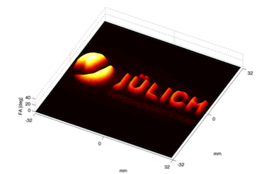
Selective excitation of an arbitrary 3D target region requires long high frequency pulses, which can be considerably shortened by means of multiple transmit channels. This project investigates new approaches for the numerically demanding calculation of the pulseshapes, in order to apply 3D selective excitation in routine applications.
Selective excitation of an arbitrary 3D target region requires long high frequency pulses, which can be considerably shortened by means of multiple transmit channels. This project investigates new approaches for the numerically demanding calculation of the pulseshapes, in order to apply 3D selective excitation in routine applications.
Group Leader
- Institute of Neurosciences and Medicine (INM)
- Medical Imaging Physics (INM-4)


