
TECHNICAL ASPECTS OF MR-PET
The hybrid scanner 3T MR-PET consists of a high-resolution BrainPET, newly constructed by Siemens, and a commercial 3T MAGNETOM Trio MRI scanner. The 9.4TMR-PET combines the BrainPET with a 9.4TMRI developed through collaboration between the Forschungszentrum Jülich and Siemens.
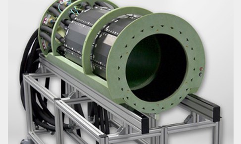
The BrainPET is a compact cylinder with a length of 72 cm and an outer diameter of 60 cm, fitting snugly into the bore of the magnet. Within its inner diameter of 32 cm, there is a birdcage head coil used for both transmitting radiofrequency pulses and receiving the signals and an inner 8-channel coil for receive only.
The BrainPET consists of 32 copper shielded cassettes, each containing six detector modules covering an axial field-of-view (FOV) of 19.2 cm. The front end of the detector module is a 12 x 12 matrix of 2.5 x 2.5 x 20 mm3 individual LSO (Lutetium oxysilicate)scintillation crystals coupled to a 3 x 3 array of avalanche photodiodes (APD).
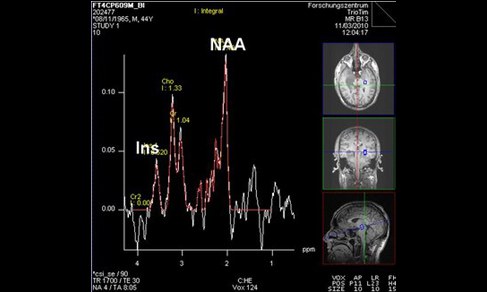
The APD are not influenced by the magnetic field and are used instead of photomultipliers which would not work in the MR environment. Since the APDs are temperature sensitive, the temperature of the BrainPET is stabilised with cooled air. The APD output signals are preamplified and transferred via long cables to the filter plate and thereafter to the data acquisition electronics. Here the coincidence lines are recorded so that PET images can be reconstructed as decribed at ( Technical Aspects of Positron-Emission-Tomography). The BrainPET offers an excellent image resolution of 3 mm at the centre.
GROUP LEADER

Univ.-Prof. Dr. Dr. h.c. N. J. Shah
Institute Director INM-4
- Institute of Neurosciences and Medicine (INM)
- Medical Imaging Physics (INM-4)
Building 15.14 /
Room 201
Room 201
+49 2461/61-6836
E-Mail

Dr. Christoph Lerche
Group Leader: PET
- Institute of Neurosciences and Medicine (INM)
- Medical Imaging Physics (INM-4)
Building 15.2v /
Room 310
Room 310
+49 2461/61-96524
E-Mail

- Institute of Neurosciences and Medicine (INM)
- Medical Imaging Physics (INM-4)
Building 15.14 /
Room 211
Room 211
+49 2461/61-6356
E-Mail
Staff
Last Modified: 09.03.2023