CONTRAST MECHANISMS
The aim of our research is to establish new methods for investigating the microscopic properties of tissue using its NMR characteristics.
MR-observable properties, for example the relaxation times of various nuclear species, are determined by different microscopic properties of the environment averaged over a distance sampled through diffusion of the nuclei during the NMR encoding time (~ 10 microns for brain tissue). The properties thus sampled and averaged include electric field gradients (for nuclei with a quadrupole moment), local magnetic field perturbations related to the distribution of different elements, motion restriction by boundaries, and concentration of specific substances (proteins, myelin, ferritin).
This is clearly a rich source of information, which has not been fully exploited until now, partly because of its complexity. For the human/mammal brain, the sensitivity of the MR relaxation times to tissue type (white matter/ grey matter) and its pathological changes (e.g. tumours) has been noticed more than 30 years ago, and the diagnostic powers of the MR contrast has revolutionized modern medicine. Learning more about the microscopic structure which gives rise to this contrast is our long-term aim.
One direction of research focuses on the measurement of different “traditional” NMR parameters (relaxation times, proton density) with high precision and accuracy using optimized methods/protocols and postprocessing methods.
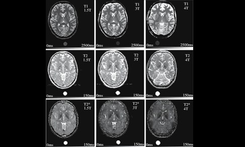
A related direction concentrates on the development of methods for quantitative imaging at very high magnetic fields. Phase and susceptibility contrast and tissue T2* contrast in high resolution imaging at very high fields are already starting to provide a deeper insight into brain structure than achievable at lower fields. Correlation of these properties with the element distribution in tissue will help clarify the origin of the contrast.
Very high-resolution imaging is a brute-force tool to characterize tissue microscopy down to the diffusion limit (~ 10 microns). We have an ongoing program of investigating different objects with very high resolution quantitative imaging and/or optimised contrast. Furthermore, we investigate possibilities for exploiting the extensive averaging required by high-resolution imaging. As a first step, a method for reconstruction of thin slices from multiple acquisition of thick slices (superresolution reconstruction) has been developed.
Projects
Origin of Phase Contrast
Measurement of the NMR frequency of water protons is one of the most sensitive ways to measure magnetic fields.
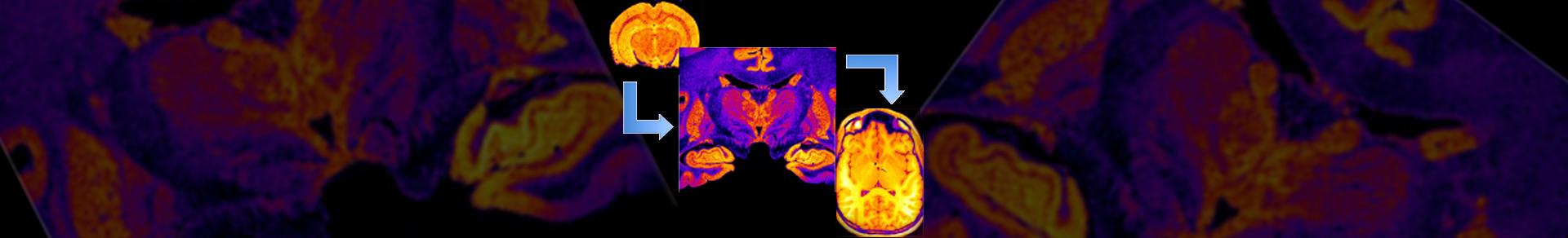
Animal Imaging
A 9.4T scanner for small animal imaging has been developed in our Institute and has taken several incarnations over the years. For the latest developments see the page of Team Hardware. Two consoles are available for imaging/spectroscopy: a Varian UnityInova console and a unique Siemens console with parallel transmit and receive capabilities.
Measurement of the NMR frequency of water protons is one of the most sensitive ways to measure magnetic fields.
High-resolution Imaging
Due to the long measurement times required, most examples are restricted to post mortem tissue or animal imaging.
Methods for Quantitative Imaging
Mapping of the water content in the brain is one of the most challenging tasks in quantitative imaging.
Due to the long measurement times required, most examples are restricted to post mortem tissue or animal imaging.
Phase Imaging and Susceptibility Reconstruction
The importance of MR phase imaging has strongly increased during the last years. The investigation of phase data enables detailed insights into microstructure and magnetic properties of tissue, in particular field information and magnetic susceptibility.
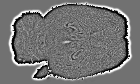
Deconvolution of Signal Decay and Recovery of T2* Information
In magnetic resonance imaging (MRI) quantitative measurement of the effective transverse relaxation time, T2*, yields to be an important contrast mechanism in manifold applications of MRI such as BOLD imaging in functional MRI, susceptibility weighting, etc.
The importance of MR phase imaging has strongly increased during the last years. The investigation of phase data enables detailed insights into microstructure and magnetic properties of tissue, in particular field information and magnetic susceptibility.
Analysis of Brain Tissue Spatial Correlation by Means of Variography
Neuro-degenerative diseases as well as normal ageing processes are often accompanied by structural changes in the brain tissue. In order to quantify and monitor spatial homogeneity and correlation of tissue from different subjects and to identify structural anisotropies of the brain, methods known from geostatistics are picked up and tailored to the analysis of MR images.
Neuro-degenerative diseases as well as normal ageing processes are often accompanied by structural changes in the brain tissue. In order to quantify and monitor spatial homogeneity and correlation of tissue from different subjects and to identify structural anisotropies of the brain, methods known from geostatistics are picked up and tailored to the analysis of MR images.
Project Leader
- Institute of Neurosciences and Medicine (INM)
- Medical Imaging Physics (INM-4)