
MR-PET
Since the end of the twentieth century, MRI and PET have revolutionised in vivo imaging, with PET providing metabolic information with high levels of sensitivity and MRI being the method of choice for structural imaging.
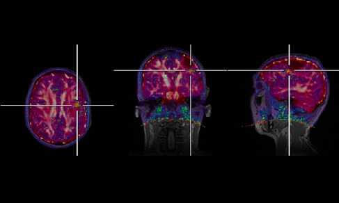
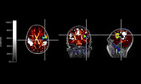
Due to the complementary nature of the information afforded by each modality, the combination of PET and MRI is advantageous to aid diagnostics and treatment planning. This can be achieved by registering and fusing images from the two modalities using appropriate software tools. However, scientists in INM4 are currently working to develop new hybrid imaging technology in the form of a scanner that allows PET and MRI images to be acquired simultaneously.
The initial steps in the development of a hybrid scanner required the development of a new type of MR-compatible PET detector, which was first realized for studying small animals. Since then, the first industrial prototypes of integrated MR-PET scanners for human brain studies were installed in 2008. Beyond time saving in patient management and the optimal spatial overlay of anatomic and functional information, this technique offers new perspectives with regard to the simultaneous observation of physiological and biochemical processes. For example, the use of simultaneous MR-PET imaging enables functional brain activation and the interaction of receptor ligands during special activation tasks or pharmacological intervention to be explored in humans. Thus, MR-PET opens new horizons in multiparametric neuroimaging, particularly in terms of the investigation of dynamic MR-PET information.
Presently, the institute holds two hybrid MR-PET scanners at high field and ultra high field with 3 Tesla and 9.4 Tesla, respectively. Both scanners are based on whole body Siemens MRI systems and are equipped with identical Siemens BrainPET inserts. The PET inserts are based on Avalanche photo diodes (APD), since conventional photo multipliers (PMT) do not work inside the magnetic field of the MR-scanners. The positioning of the BrainPET insert in the centre of the magnet offers the opportunity for iso-centred imaging in space and time.


Group Leader

Univ.-Prof. Dr. Dr. h.c. N. J. Shah
Institute Director INM-4
- Institute of Neurosciences and Medicine (INM)
- Medical Imaging Physics (INM-4)
Building 15.14 /
Room 201
Room 201
+49 2461/61-6836
E-Mail
Dr. Christoph Lerche
Group Leader: PET
- Institute of Neurosciences and Medicine (INM)
- Medical Imaging Physics (INM-4)
Building 15.2v /
Room 310
Room 310
+49 2461/61-96524
E-Mail

- Institute of Neurosciences and Medicine (INM)
- Medical Imaging Physics (INM-4)
Building 15.14 /
Room 211
Room 211
+49 2461/61-6356
E-Mail
Staff

Priv.-Doz. Dr. Philipp LohmannGroup Leader: Digital Translational NeuroimagingBuilding 15.2 / Room 201a+49 2461/61-96357

Last Modified: 06.03.2023