Nanodiscs as first-class model membranes
Nanodiscs ermöglichen das Studium von Membranproteinen
Nanodiscs are self-assembled model membranes that resemble high density lipoprotein (HDL) particles. They consist of a small lipid bilayer patch and two copies of an amphipathic membrane scaffold protein (MSP) that shield the hydrophobic rim of the membrane fragment from water. Different scaffold proteins are in use, most popular are recombinantly produced variants of human apolipoprotein A-I. Nanodiscs have been optimized, promoted, and characterized in the laboratory of Stephen G. Sligar at the University of Illinois at Urbana-Champaign.

Nanodiscs afford solubilization of membrane proteins in a functional state, thus making them available for biophysical analysis techniques previously restricted to soluble proteins.
Nanodiscs in solution NMR studies
We demonstrated that integral membrane proteins reconstituted in nanodiscs can be studied by high resolution liquid state NMR. An isotope labeled polypeptide containing the transmembrane and cytoplasmic domains of human CD4 was incorporated into nanodiscs. Two-dimensional 1H, 13C HSQC NMR spectra show correlation signals of both the 13C-labeled CD4 and the lipid molecules (13C at natural abundance).
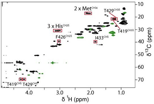
Glück et al. (2009) J Am Chem Soc 131: 12060-61
Nanodiscs in surface plasmon resonance studies
Nanodisc-reconstituted integral membrane proteins can serve as analyte in SPR studies. We inserted a fusion of the transmembrane domain of CD4 and ubiquitin carrying a N-terminal decahistidine tag into nanodiscs. Binding of the nanodisc-inserted fusion protein to an immobilized antihistidine monoclonal antibody was quantified in SPR experiments.
