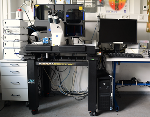
Live cell imaging setup

Unlike immunofluorescence of fixed samples, life cell imaging focuses on living cells. Imaging a living cell is by definition minimally invasive imaging. As long as phototoxicity is averted, humidity, and CO2 concentration are in a physiological regime cells can be imaged for a long period. Thus we are able to investigate cellular growth and the influence of structural confinements on the outgrowth is of particular interest in our institute.
As imaging methods we can use bright field, phase contrast, or differential interference contrast microscopy as well as fluorescent microscopy, while we image from a single spot, multiple spots or regions on our substrate. By using fluorescently labeled proteins we are able to investigate the influence from patterned protein on the cellular development on a cellular level.
Beside these applications the setup (Axio Observer.Z1, Zeiss) is equipped with additional features such as a high speed and light sensitive PCO-camera system and a FRAP (Rapp OptoElectronic) unit with a 437 nm laser. The fast camera enables us to observe cellular activity with single cell resolution and a temporal resolution of several milliseconds on large substrates (1.248mmx0.666mm).
In addition to these functions the whole variety of an inverted microscope equipped with a fluorescence light source is available. Applications ranging from single images with different fluorescence channels to tile regions acquired in a z-stack
Contact:
Dr. Vanessa Maybeck
Tel.: +49-2461-61-3675
e-mail: v.maybeck@fz-juelich.de
