2020-11--Viviana.normal.png
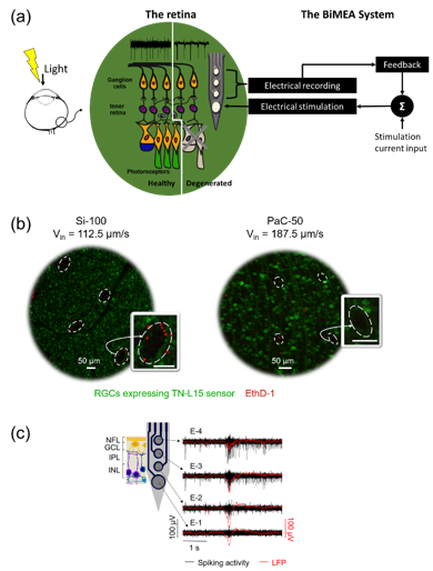
(a) Sketch of the BiMEA strategy, (b) dead cell stainings after intraretinal insertions in TN-L15 mouse retinas (red: dead cells; green: live retinal ganglion cells, and (c) intraretinal recordings of a generated rd10 mouse retina captured by a fully inserted parylene-C shank (50µm wide, 180 µm long), black and red traces show spiking activity of the retina and local field potentials, respectively.
Last Modified: 27.09.2021