Cryo-EM methods
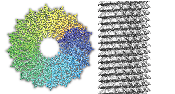
The cryo-EM method has become very powerful in the past several years and for many structural biologists it is becoming the method of choice. The full potential of the technique, however, is still to be realized as the scientific community is in great need of hardware-based and software improvements. Although the first 3D images of biological specimens at true atomic resolution have been obtained recently (Fromm et al., 2015, Weis et al., 2019), the routine resolution of cryo-EM still lags behind in detail and quality in comparison with common material science electron microscopy as it is imaged at the Ernst-Ruska Centre (ER-C) 1 and 2. Therefore, in collaboration with the ER-C-1 and ER-C-2 we will apply new imaging hardware to biological samples. Our technique development also includes novel sample preparation, data acquisition and image processing methods that we benchmark using biological test specimens. One particular focus of our activities is the development and application of cryo-scanning transmission electron microscopy that we study with collaborators Knut Müller-Caspary and Henning Stahlberg in the framework of an ERC synergy grant. We showed that for plunge-frozen test specimens, we can elucidate the three-dimensional structures at near-atomic and subnanometer resolution (Lazic et al., 2022; Küçükoğlu et al. 2025). We apply a series of innovative cryo-EM methods to the structures of challenging biological systems with a particular focus on membrane assemblies.
Related Publications
- Küçükoğlu, B., Mohammed, I., Guerrero-Ferreira, R.C., Ribet, S.M., Varnavides, G., Leidl, M.L., Lau, K., Nazarov, S., Myasnikov, A., Kube, M., Radecke, J., Sachse, C., Müller-Caspary, K., Ophus, C., Stahlberg, H., 2024. Low-dose cryo-electron ptychography of proteins at sub-nanometer resolution. Nat Commun 15, 8062.
- Lazić, I., Wirix, M., Leidl, M.L., de Haas, F., Mann, D., Beckers, M., Pechnikova, E.V., Müller-Caspary, K., Egoavil, R., Bosch, E.G.T., Sachse, C., 2022. Single-particle cryo-EM structures from iDPC-STEM at near-atomic resolution. Nat Methods 19, 1126–1136.
- Weis, F., Beckers, M., Hocht, I., Sachse, C., 2019. Elucidation of the viral disassembly switch of tobacco mosaic virus. EMBO Rep 20.
- Fromm, S.A., Bharat, T.A.M., Jakobi, A.J., Hagen, W.J.H., Sachse, C., 2015. Seeing tobacco mosaic virus through direct electron detectors. J. Struct. Biol. 189, 87–97.