Polymer Nanopillars Induce Neuronal Adhesion and Axon Growth
Polymer Nanopillars Induce Increased Paxillin Adhesion Assembly and Promote Axon Growth in Primary Cortical Neurons. - F. Milos, A. Belu, D. Mayer, V. Maybeck, A. Offenhäusser, Adv. Biology 2021, 5, 2000248.
The complexity of the extracellular matrix consists of micro- and nanoscale structures that influence neuronal development through contact guidance.
Substrates with defined topographic cues mimic the complex extracellular environment and can improve the interface between cells and biomedical
devices as well as potentially serve as tissue engineering scaffolds. This study investigates axon development and growth of primary cortical neurons on OrmoComp nanopillars of various dimensions. Neuronal somas and neurites form adhesions and F-actin accumulations around the pillars indicating a close contact to the topography. In addition, higher pillars (400 nm) confine the growing neurites, resulting in greater neurite alignment to the topographical pattern compared to lower pillars (100 nm). A comprehensive analysis of growth cone dynamics during axon development shows that nanopillars induce earlier axon establishment and change the periodicity of growth cone dynamics by promoting elongation. This results in longer axons compared to the flat substrate. Finally, the increase in surface area available for growth cone coupling provided by nanopillar sidewalls is correlated with increased assembly of paxillin-rich point contact adhesions and a reduction in actin retrograde flow rates allowing for accelerated and persistent neurite outgrowth.
In this study, the authors used nanopillar arrays to investigate the
effects of isotropic topography on adhesion and early devel-
opment of primary cortical neurons in vitro. A biocompatible
polymer OrmoComp was employed to fabricate frustum-shaped
nanoscale pillar arrays resulting in a transparent in vitro plat-
form compatible with conventional optical microscopy. We eval-
uated both neuronal soma adhesion and neurite outgrowth on
nanopillar arrays in comparison to flat OrmoComp substrates.
Time-lapse microscopy was used to systematically study axonal
growth dynamics, including the time until axon establishment,
periodicity of growth phases (elongation, pausing, retraction),
and the resulting axon length. Finally, the influence of pillar
topographies on point contact adhesions and F-actin retrograde
flow in the growth cone was analyzed.
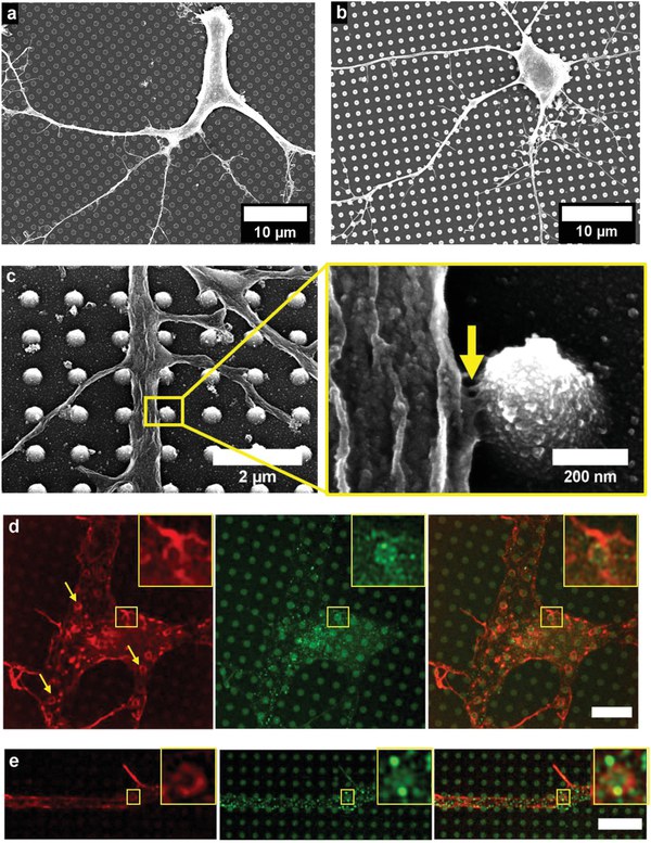
FIGURE 1. Neuronal growth and adhesion on nanopillar arrays. Neurites extended a) on and between the pillars on L-arrays (100 nm high pillars), while on H-arrays (400 nm high pillars), neurites were mostly confined to the space in b) between the pillars and c) (arrow) adhered to pillar sidewalls. d,e) Nanopillars perturbed the actin cytoskeleton (red) visible by the formation of ring-like structures around the pillars (arrows). These structures were often localized with paxillin-rich point contacts (green; zoomed-in inset). Scale bars: 5 µm.
Publication: Frano Milos, Andreea Belu, Dirk Mayer, Vanessa Maybeck, Andreas Offenhäusser, Polymer Nanopillars Induce Increased Paxillin Adhesion Assembly and Promote Axon Growth in Primary Cortical Neurons, 14 January 2021, Adv. Biology 2021, 5, 2000248. (Pub. by Wiley-VCH GmbH.) DOI: https://doi.org/10.1002/adbi.202000248
Contact:
Prof. Dr. Andreas Offenhäusser
Institute of Biological Information Processing-Bioelectronics (IBI-3)
Tel.: +49 2461 61-2330
E-Mail: a.offenhaeusser@fz-juelich.de

