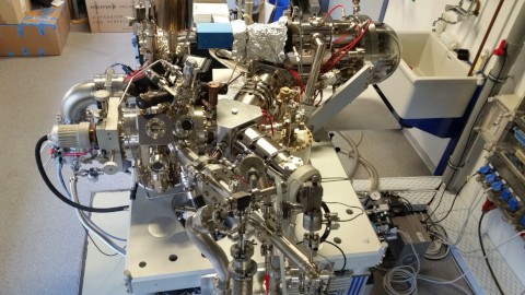
Aberration-Corrected Spectromicroscopy
We are running an aberration-corrected spectroscopic low energy electron microscope (Elmitec AC-SPE-LEEM) for real- and k-space imaging of surfaces and thin films. The microscope is one of the best of its kind in terms of spatial resolution and transmission. We use it for the observation of kinetic processes at surfaces in real time.
In LEEM mode, our microscope images the structure and morphology of surfaces with a spatial resolution better than 2 nm. The images are formed by low-energy electrons that are elastically backscattered from the sample surface. The LEEM mode offers a broad variety of contrast mechanisms, e.g. amplitude and phase contrast, bright and dark field microscopy, work function or chemical contrast. This allows the study of, e.g., surface reconstructions, growth processes and self-organisation at surfaces, phase transitions, and other kinetic processes. As an example, a movie below shows a series of phase transitions in a mixed molecular monolayer film [1, 2]. We also investigated 2D materials using LEEM, in particular hexagonal boron nitride (hBN) [3, 4, 5].
To improve the utility of the instrument even further, we have developed a method of linear exit wave reconstruction from a through-focal series of LEEM images. It can be used to enhance the resolution (by exploiting the full information limit of the microscope) and the image contrast (by considering the electron phase shifts) [6].
Because LEEM is a diffraction-based imaging technique, the microscope can easily (and quickly) be switched to k-space imaging, simply by projecting a diffraction plane onto the channel plate detector instead of an image plane. The microscope then records (micro-) low energy electron diffraction (µLEED) images, useful for identifying the crystallographic structure from surface areas down to approx. 1 µm in diameter.
When using a UV photon source instead of electrons for illuminating the sample, photoemitted electrons can be detected by the microscope as well (photoemission electron microscopy or PEEM). Our microscope is equipped with a high-flux He-lamp (Focus HIS-14 HD) and a hemispherical electron energy filter, allowing for spectroscopic investigations as, e.g., three-dimensional k-space mapping (momentum microscopy, see movie below) and photoemission tomography [7].

