Diffraction Methods and Electron Microscopy
The banner image shows a LEEM image of a 0°-rotated monolayer graphene on a 6H-SiC(0001) substrate prepared via the surfactant-mediated growth method.
About
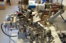
Diffraction methods provide quantitative structural information of periodic structures and are therefore complementary to scanning probe microscopies. We employ both, electron and x-ray diffraction, to study molecular and quantum materials. Our aberration-corrected electron spectromicroscope plays a special role here, as it combines nanometer-resolved microscopy with micro-diffraction and spectroscopic capabilities.
Research Topics
- Aberration-corrected spectromicroscopy
- Chemically-resolved vertical structures with sub-Angstrom resolution
- Interfaces between molecules and crystalline phases
- Epitaxial 2D materials
Members
Monja StettnerPhD student at Peter Grünberg Institute (PGI-3) Building 02.4w / Room 128+49 2461/61-6313
Research
Recent Publications
Last Modified: 20.01.2026


