de en
Jülich Supercomputing Centre (JSC)
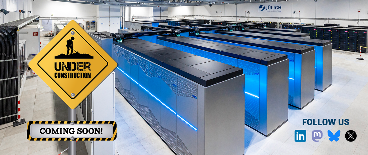
The page is currently under construction. All information can still be found on: https://www.fz-juelich.de/en/ias/jsc
The page is currently under construction. All information can still be found on: https://www.fz-juelich.de/en/ias/jsc