“It’s as if a door to a new world has been opened,” says Privatdozent Dr. Gregor Hagelüken: “When I see the structure of a protein in front of me in all its detail for the first time, it often helps me to understand how this molecular machine works.”
Protein molecules perform a wide variety of tasks in the human body. The structural biologist from the University of Bonn aims to understand how the three-dimensional shape of proteins relates to their functions. Hagelüken is particularly interested in molecules that play a role in the immune system: “I see potential applications in cancer therapy, for example, or in the search for alternatives to antibiotics.”
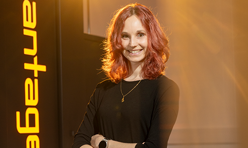
Penghan Lu has been working at Forschungszentrum Jülich for 11 years. He particularly values the international working environment and the collaboration with industry partners.Copyright: Forschungszentrum Jülich / Sascha Kreklau
He visualizes the structure of proteins using cryo-electron microscopy, a specialized form of electron microscopy. In many cases, it delivers results much faster than the older method of X-ray crystallography. However, the problem lies in the availability of equipment, says Hagelüken: “There are only a few sites in Germany.” One of these is Jülich’s Ernst Ruska-Centre (ER-C), which offers electron microscopes that are unique worldwide.
For Hagelüken, the ER-C is an ideal fit for several reasons – its proximity to Bonn, the fact that there are no costs for the universities using the facility, and the on-site expertise: “My team and I initially received comprehensive training on the equipment there. The experts at Jülich showed us how to prepare the samples and assess their quality. And they carried out the first measurements together with us.” The measurements performed at ER-C have since resulted in the first publications by Hagelüken and his team.
Standard developed
At ER-C, research is carried out not only with the instruments but also on them. “We are continuing to develop the electron microscopes and methods ourselves in order to make the tiniest atomic structures visible,” explains physicist Penghan Lu. This has a long tradition. At ER-C, for example, the method of spherical aberration correction was co-invented to improve imaging. This involves compensating for unavoidable imaging errors in electron optics using complex magnetic lenses. Today, this technique is a standard feature of high-performance electron microscopes.
Penghan Lu is investigating how the resolution and contrast of electron microscopes can be increased even further, even when they are used to make fragile structures visible: “Biological samples in particular, or certain structures in materials science, are damaged by the electron beam. We are therefore developing new imaging techniques that enhance image contrast while simultaneously reducing the electron dose.”
For Gregor Hagelüken, electron microscopes are indispensable tools for his research.Copyright: Johann F. Saba/UKB
As a national research infrastructure, the centre has been breaking new ground since 2019 under the project name ER-C 2.0 – with innovative equipment and applications for science and industry. For example, the researchers are developing instruments that combine electron microscopy with other techniques, such as atom probe tomography. Lu: “Materials scientists often use these complementary techniques to determine the three-dimensional chemical distribution and atomic structure of a sample. But to do so, they have to transfer the sample from one instrument to another. Our combined instrument eliminates this step.
The experts at Jülich are also working on electron microscopes in which they can manipulate samples in a targeted way, for example by allowing the material to grow into microstructures under the microscope and continuously monitoring the process. “This presents us with entirely new challenges. Among other things, we need to significantly improve the vacuum in our instruments. To do so, we are working closely with manufacturers,” says the physicist.
The unique centre attracts researchers from every continent. “Such collaborations often develop into long-term partnerships, creating new synergies between the centre and research institutions around the world,” says Lu.
How it began In 1990, something unique in Europe was established at Jülich: a centre for high-resolution electron microscopy with four state-of-the-art instruments. At the time, the only comparable facilities existed in Japan and the USA. Electron microscopes allow materials to be characterized down to the atomic scale. In the 1990s, experts from Darmstadt, Heidelberg, and Jülich developed aberration correction, a method that compensates for physically induced imaging errors and thus enables higher resolutions. In 2004, Jülich, together with RWTH Aachen University, founded the Ernst Ruska-Centre (ER-C), a national centre of excellence for ultra-high-resolution electron microscopy. The centre’s infrastructure is currently being expanded as part of the ER-C 2.0 project.
70 Years of Forschungszentrum Jülich – Our History
For 70 years, the Forschungszentrum Jülich has been tackling the big questions of our time. From energy research and supercomputing to AI and the sustainable circular economy: this journey through time highlights the milestones on the path to becoming one of Europe’s largest interdisciplinary research centres.