Glimpse under the Bonnet of Complex Molecules
Individual atoms as contrast media for scanning tunnelling microscopes
Jülich, 5 October 2011 – Scanning tunnelling microscopes are among the most important and most widely used tools for visualizing structures at the atomic level. In the past, however, it was virtually impossible to use them to penetrate inside complex molecules. Jülich researchers have now cleared another hurdle in order to overcome this limitation. Using individual atoms between the tip of the microscope and the sample as a sort of contrast medium, they image the inner structure of the molecule as well as the intermolecular forces. The method was presented on 3 October 2011 in the Journal of the American Chemical Society (DOI: 10.1021/ja204626g).
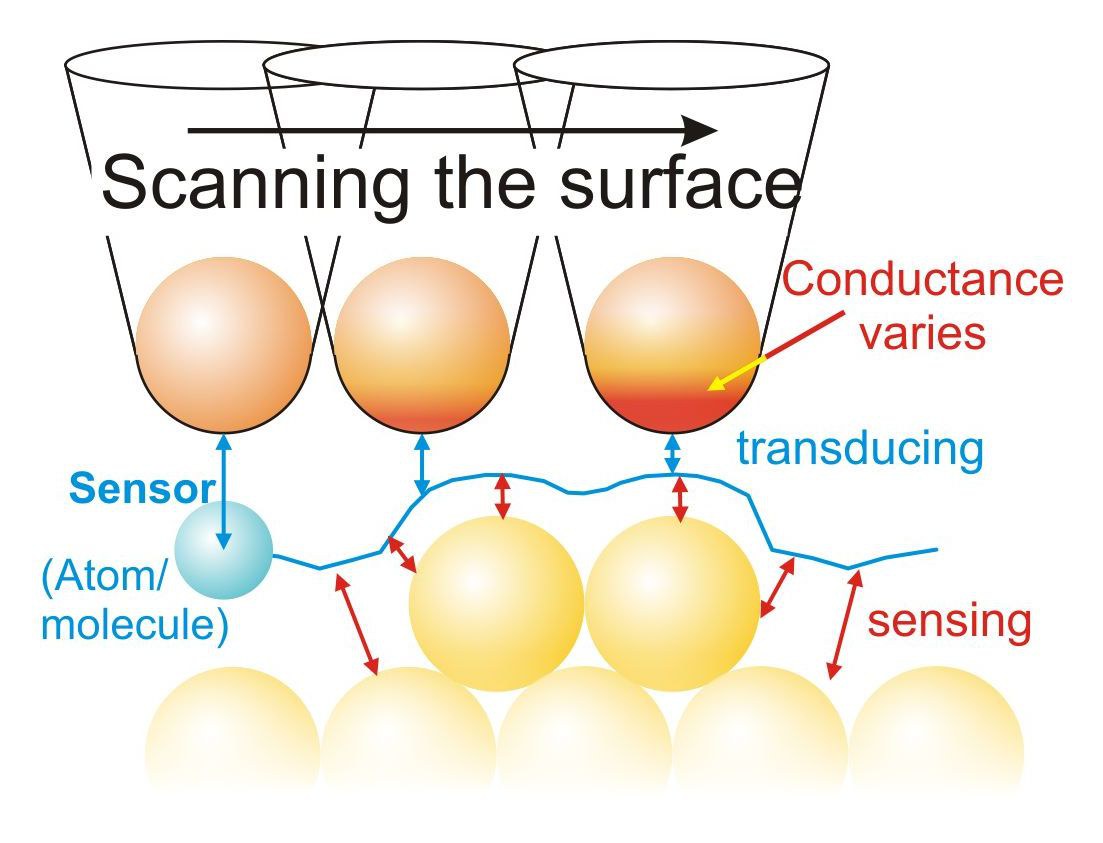
Biomolecules, proteins or organic semiconductors for future electronic components – many of the materials at the heart of the nanosciences proved difficult to investigate with conventional scanning tunnelling microscopes in the past. These microscopes trace the surface of a sample using a fine metal tip that is often tapered to a single atom, and in doing so, they determine the strength of an electrical current. However, this "tunnelling current" only detects the external electron shell. As these shells stretch the length of the entire molecule in the case of many complex molecules, the conventional use of such microscopes does not provide any indications of the deeper atomic molecular structure.
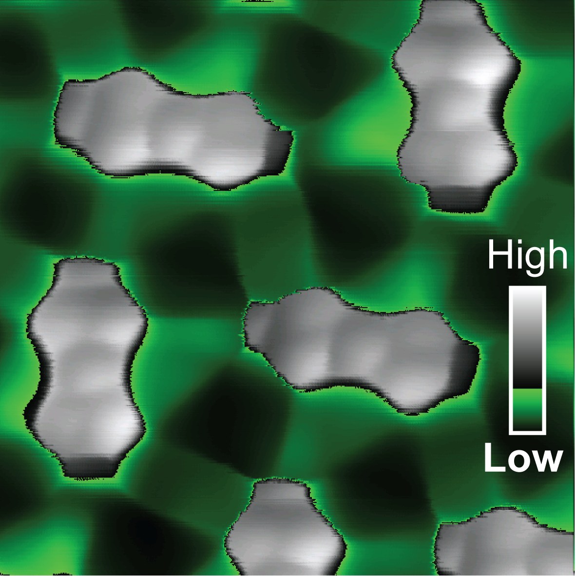
The Jülich group headed by Dr. Ruslan Temirov and Prof. Stefan Tautz from the Peter Grünberg Institute have been investigating methods of expanding the potential of scanning tunnelling microscopy for a number of years. In their article, they describe how different types of atoms or molecules can be used as signal transducers with different characteristics. The atoms are attached to the tip of the microscope. Small displacements cause them to react extremely sensitively to the contours of molecules, which in turn influences the measurable tunnelling current. In this way, conventional, industrially manufactured scanning tunnelling microscopes can be used to produce images of the order of atoms inside complex molecules and even to visualize intermolecular forces, such as hydrogen bridge bonds.
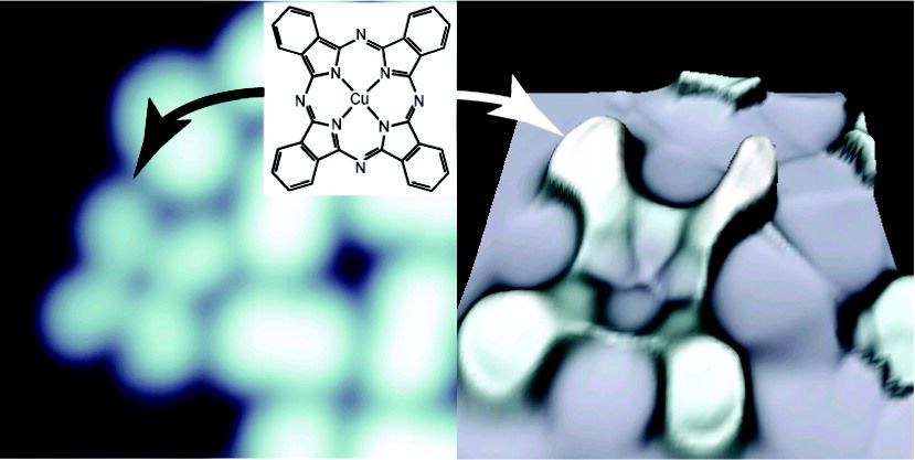
"The surprising thing about this method is that it is so amazingly simple. By using different types of atoms, we will be able to make customized contrast media available for various applications in no time at all," says Ruslan Temirov. The experiments at Jülich used xenon, carbon monoxide and methane, each of which behaves with varying levels of sensitivity, making them appropriate for different ranges. "This work represents the first step towards a standard application. It is conceivable that very different materials will be used as sensors in the future," says Temirov.
While working on developing this method, the researchers published the first images of the inner structure of complex organic molecules back in 2008. A patent has also been filed for the method of attaching individual atoms or molecules to the tip of a scanning tunnelling microscope, which then act as sensors transducing signals from the sample surface. Originally, heavy hydrogen molecules were used as sensors, but these could not be precisely dosed. Hydrogen moves very readily and is therefore rather restless. Furthermore, it is also almost invisible in the scanning tunnelling microscope. "The previous method did not allow us to identify how many molecules were located between the probe head and the sample. You have to imagine the hydrogen as a liquid that spreads out over the entire sample," says Jülich researcher Dr. Christian Wagner. Having explained the basic physical principles upon which the new technology is based around a year ago, the researchers have now succeeded in taking another step towards establishing a broad range of applications.
Original publication:
Single Molecule and Single Atom Sensors for Atomic Resolution Imaging of Chemically Complex Surfaces
Georgy Kichin, Christian Weiss, Christian Wagner, F. Stefan Tautz, Ruslan Temirov
J. Am. Chem. Soc.
DOI: 10.1021/ja204624g
Publication date (online): 3 October 2011
Direct link to the online publication:
http://pubs.acs.org/doi/abs/10.1021/ja204624g
Further information:
How a scanning tunnelling microscope works
Research at the Peter Grünberg Institute
Press release from 20.10.2010 "Jülich Researchers Take a Look Inside Molecules"
Article in Welt der Physik from 30.08.2010, "Mikroskop macht Atomstruktur von Molekülen sichtbar"
Press release from 27.02.2009, "Latest Issue of 'Science': Nano-Sonar Uses Electrons to Measure under the Surface"
Contacts:
Prof. Stefan Tautz
s.tautz@fz-juelich.de
Tel.: +49 2461 61 4561
Dr. Ruslan Temirov
r.temirov@fz-juelich.de
Tel.: +49 2461 61 3462
Dr. Christian Wagner
c.wagner@fz-juelich.de
Tel.: +49 2461 61 3538
Press Contact:
Tobias Schlößer
Tel.: +49 2461 61 4771
t.schloesser@fz-juelich.de