Inside a tumor: Purple bacteria visualize ‘big eaters’
Düsseldorf / Jülich / Munich, 13 March -- Cancer presents an ongoing challenge for physicians, particularly in the diagnosis and treatment of the disease. This is, amongst other things, due to tumor heterogeneity. A team of scientists from the Helmholtz Zentrum München, the Jülich Research Center, the Technical University of Munich and the Heinrich Heine University Düsseldorf has now shown that harmless purple bacteria of the genus Rhodobacter are capable of visualizing aspects of this heterogeneities in the tumors. With the aid of optoacoustic imaging, the researchers used these microorganisms to visualize cells of the immune system, so-called macrophages (Greek for ‘big eaters’), that also play a role in tumor development. Their research findings have now been published in Nature Communications.
Many cancers form solid tumors. Inside, such tumors reveal major differences at the cellular and molecular level. One of these concerns the localization and activity of macrophages. Although these cells are essential for a healthy immune system, they also play a key role in tumor development as they are involved in unwanted inflammation of the tumor tissue – a process that leads to the further progression of the disease. With the aid of photosynthetic bacteria, new optoacoustic techniques, which indicate where such macrophages are present and active, have now been developed.
“We were able to demonstrate that bacteria of the genus Rhodobacter, which are harmless to humans, are suitable as indirect markers of macrophage presence and activity,” says Dr. Andre C. Stiel, head of the Cell Engineering Group at the Institute of Biological and Medical Imaging (IBMI), Helmholtz Zentrum München. Rhodobacter occurs worldwide in stagnant and flowing water. The bacteria produce different pigments in order to carry out photosynthesis. These include bacteriochlorophyll a, which is suitable for multispectral optoacoustic tomography (MSOT) examinations to localize solid tumors.
During a MSOT scan, light is initially converted into sound, then into visual information. Initially, a weak, pulsating laser beam is directed towards the body. When the beam encounters molecules and cells, they heat up minimally and respond with minimal vibrations, which in turn generate acoustic signals. These are then picked up by sensors and converted into images. The way in which the individual cells and molecules react to the laser depends on their optical properties – in this case, for example, on the properties of bacterial pigments.
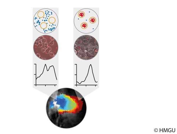
How does the principle work? Macrophages engulf bacteria as part of their natural scavenging activity, which is known as phagocytosis. This alters the surroundings of the bacteria, their absorption of electromagnetic radiation and, as a result, also the optoacoustic signal. Rhodobacter bacteria thus act like sensors for scientists, providing them with information about the presence and activity of macrophages.
"In further steps, these bacteria will enable novel approaches to non-invasive technologies and so open up entirely new possibilities for innovative diagnostic and therapeutic procedures,” adds Dr. Thomas Drepper, who heads the Bacterial Photobiotechnology Group at Heinrich Heine University Düsseldorf. In future, bacteria may be able to reveal the location of a tumor and also detect increased macrophage activity. Depending on their localization, the macrophages could provide information about unwelcome inflammations or the desired response to immunotherapies, and could ultimately be used to improve treatments strategies.
Original publication:
Lena Peters et al. (2019): Phototrophic purple bacteria as optoacoustic in vivo reporters of macrophage activity. Nature Communications, DOI: 10.1038/s41467-019-09081-5
Further information
http://www.iet.uni-duesseldorf.de/
Contacts:
Dr. Thomas Drepper
Institute of Molecular Enzyme Technology (IMET)
Forschungszentrum Jülich
Tel.: +49 2461 61-4173
E-Mail: t.drepper@fz-juelich.de
Press contact
Annette Stettien
Corporate Communications
Forschungszentrum Jülich
+49 2461 61-2388
E-Mail: a.stettien@fz-juelich.de