Microscopic brain map: ComSLI makes networks of nerve fibers visible in differently prepared tissue sections
Microscopic brain map: ComSLI makes networks of nerve fibers visible in differently prepared tissue sections
An international team of researchers has successfully applied the recently developed imaging technique ComSLI (Computational Scattered Light Imaging) to brain sections prepared using a wide range of methods, enabling the visualization of the brain’s complex fiber network with micrometer precision. This achievement marks an important advance that opens new possibilities for neuroscience and biomedical research—such as renewed, in-depth analyses of existing tissue sections. Their findings have now been published in the prestigious journal Nature Communications.
Accurate mapping of nerve fibers in the brain is essential for understanding its functions and potential disorders. However, capturing this intricate network of neural connections across large regions with high precision has long posed significant challenges. The ComSLI technique enables the visualization of the direction and trajectories of complex fiber structures in the brain with micrometer-level resolution. In this approach, ultrathin brain sections are illuminated from multiple directions, and the light scattered perpendicularly is captured using a high-resolution camera. The scattering patterns reveal the orientation and crossings of the underlying fibers.
Researchers from Stanford, Delft, Rotterdam, and the Institute of Neuroscience and Medicine (INM-1) in Jülich have now significantly extended the applicability of ComSLI. Their study demonstrates that the technique can map fiber tract networks in various histological preparations—independently of the sample treatment method—with micrometer precision. This includes cell-body-stained whole-brain sections, tissues embedded using different techniques, and even unstained or fresh samples—an achievement not previously possible. Some of the brain sections analyzed originated from the INM-1 in Jülich and the Cécile and Oskar Vogt Institute of Brain Research in Düsseldorf.
The study builds upon expertise in 3D Polarized Light Imaging (3D PLI) , which for the first time made it possible to visualize the fine fiber-tract architecture of brain tissue in three dimensions.
The new method now allows researchers to extract additional information from existing series of tissue sections. For example, in one sample from a second “BigBrain” dataset, fiber pathways were visualized alongside previously mapped cell-body distributions. Similar to its predecessor—which constitutes a central element of the European research infrastructure EBRAINS—this second “BigBrain” represents a three-dimensional reconstruction of a complete human brain derived from several thousand cell-body-stained serial sections.
Beyond fundamental research, this approach also holds promise for medical diagnostics. The researchers were able to detect tissue alterations associated with diseases such as Multiple sclerosis and leukoencephalopathy, visualize damaged fiber tracts in regions including the hippocampus, and demonstrate the advantages of this method over conventional techniques used in Alzheimer’s disease investigations.
The study went one step further: in addition to brain tissue, the team also mapped fiber structures in other tissue types, such as muscle, bone, and blood vessels. In each case, the finest fiber arrangements became visible, providing insights into their functional organization. These findings underline that the potential of ComSLI extends far beyond brain research, offering powerful new tools for studying a wide variety of organs and tissue types and for elucidating pathological processes.
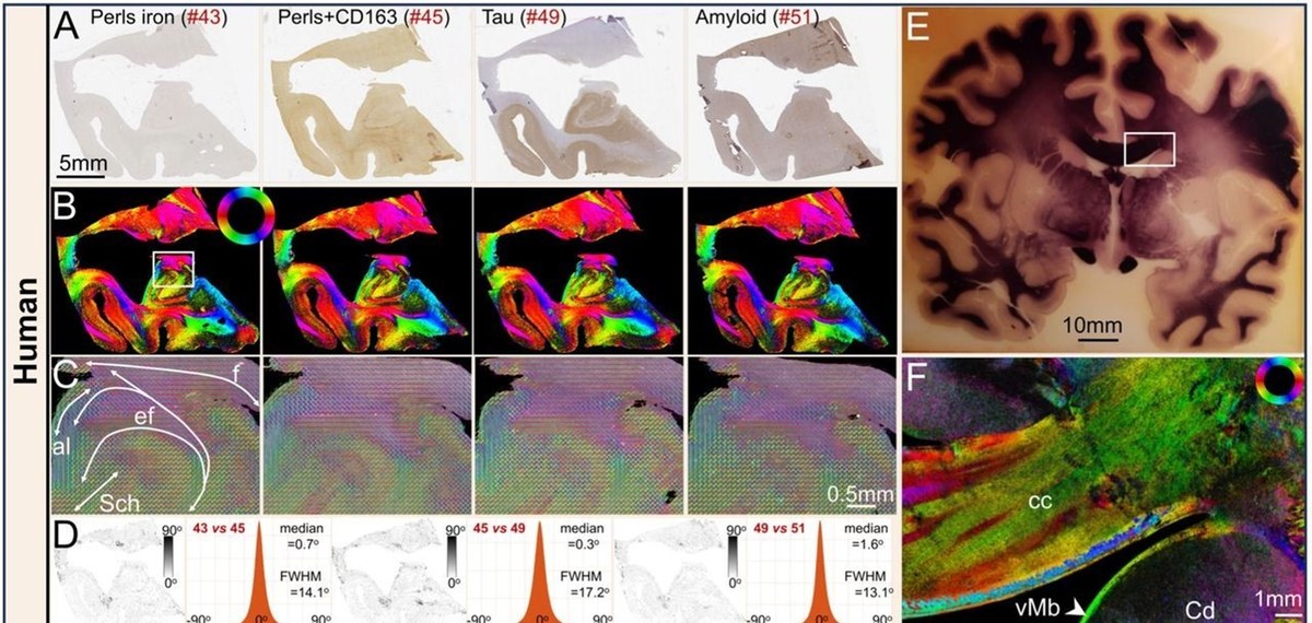
ComSLI can detect the orientation of nerve fibers independently of sample preparation or species. The figure shows examined brain sections from differently stained human hippocampi (A) and their representation using ComSLI (B). Even in an approximately 120-year-old myelin-stained human brain section (E), fiber tracts in the marked area could be visualized with ComSLI." | Copyright: Georgiadis M et al., Micron-resolution fiber mapping in histology independent of sample preparation. Nat. Comm. (…..)
Original Publication
Georgiadis M, von der Heiden F, Abbasi H, Ettema L, Nirschl J, Taghavi HM, Wakatsuki M, Liu A, Ho WHD, Carlson M, Doukas M, Koppes SA, Keereweer S, Sobel RA, Setsompop K, Liao C, Amunts K, Axer M, Zeineh M, Menzel M. Micron-resolution fiber mapping in histology independent of sample preparation. Nat Commun16, 9572 (2025). https://doi.org/10.1038/s41467-025-64896-9