Figure 1.Exemplary bode plot showing the magnitude of the electrochemical impedance spectra of PEDOT:PSS electrodes. These electrodes exhibit impedances below 100 kΩ at 1 kHz, a frequency relevant for recording neuronal spikes.
To ensure optimal functionality, we rigorously evaluate novel neurotechnologies through a series of pre-clinical tests, including in vitro, ex vivo, cadaveric, and functional in vivo studies. We assess both the physical properties of our devices (using electrochemical, mechanical, and optical methods) and their functional performance in both acute and chronic settings.
Electrochemical characterization includes techniques such as impedance spectroscopy, cyclic voltammetry, and voltage transient analysis in response to electrical stimuli. While miniaturized electrodes, typically 10-30 µm in size, are desirable to match the dimensions of neuronal somas and enable single-cell sampling resolution, there is often a trade-off between electrode size and electrochemical performance. As electrode size decreases, impedance, which reflects the resistance to current flow, increases, affecting the ability of the electrodes to record and stimulate neural activity across a wide range of frequencies. Therefore, we aim to balance electrode dimensions and electrochemical performance by characterizing and implementing different strategies, such as the electrodeposition of metal coatings or the electropolymerization of conductive polymer coatings.
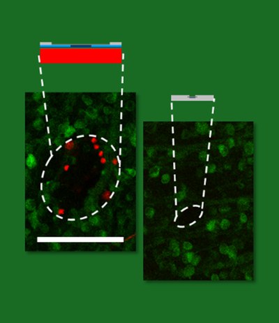
Figure 2. Exemplary maximum intensity projection of live/dead staining of explanted rodent retinas after the acute insertion of penetrating silicon- (left) and polymeric (right)-based probes. In red, dead cells stained with ethidium homodimer. In green, retinal ganglion cells expressing the calcium sensor TN-L15. Scale bar is 100 µm.
Mechanical characterization involves studying both the intrinsic properties of the materials used (e.g., Young’s modulus) and the impact of designs and fabrication methods on the mechanical stability of the devices. Additionally, we investigate the expected mechanical behaviour of our devices during implantation using analytical models, finite element method simulations, and experimental approaches, such as measuring the insertion forces or implantation feasibility using tissue phantoms and explanted or cadaveric neural models. To ensure long-term performance, we conduct accelerated ageing tests to identify potential failure modes that may arise from prolonged implantation.
To improve the integration of implantable stealth neurotechnologies in the body, we investigate the biological effects of materials, designs, and implantation methods. This includes acute and chronic studies using electrophysiology, fluorescent dyes, and immunohistochemistry to evaluate the vitality and integrity of nervous tissue. These studies allow us to evaluate the implantation footprint and examine foreign body reactions in vivo.
Abu Shihada, J. et al. Highly Customizable 3D Microelectrode Arrays for In Vitro and In Vivo Neuronal Tissue Recordings. Advanced Science11, 2305944 (2024).
Koschinski, L. et al. Validation of transparent and flexible neural implants for simultaneous electrophysiology, functional imaging, and optogenetics. J. Mater. Chem. B11, 9639–9657 (2023).
Rincón Montes, V. et al. Development and in vitro validation of flexible intraretinal probes. Sci Rep10, 19836 (2020).