Koschinski, L. et al. Validation of transparent and flexible neural implants for simultaneous electrophysiology, functional imaging, and optogenetics. J. Mater. Chem. B 11, 9639–9657 (2023).
Multimodal Neurotechnology
We explore and advance methods for monitoring and modulating neuronal activity using diverse physical modalities, including electrical, optical, and chemical approaches. To achieve multimodality, we integrate technologies and expertise in collaboration with different research groups at IBI-3.
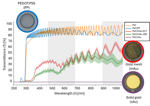
By leveraging the electrical, electrochemical, and optical properties of insulating and conducting materials, we integrate different materials to combine the strengths of electrical and optical methods for reading neural activity. While electrical methods, such as electrophysiology, allow the recording of fast-spiking activity from single neurons or low-frequency signals from groups of neurons, neuroimaging methods, such as widefield and two-photon imaging, allow capturing neuronal calcium dynamics with cell-type specificity over neuronal regions or with single-cell resolution, respectively. For example, materials such as PEDOT:PSS exhibit a high degree of transparency across a wide range of wavelengths, making them suitable for use in combination with neuroimaging methods and optogenetic stimulation.

To enable multimodal neuronal recordings, we develop transparent microelectrode arrays designed to be resistant to photo artifacts and provide long-term stability, facilitating the integration of electro- and opto-physiology in chronic in vivo neural applications. Hence, functional mapping of neural responses across brain regions correlating calcium dynamics with electrical signals is possible.
References
Last Modified: 25.03.2025