Kirigami-inspired folds for brain research: Jülich scientists create novel 3D neuroprobes
Kirigami-inspired folds for brain research: Jülich scientists create novel 3D neuroprobes
12 June 2025
Copyrights: Adobe Stock
A research team from the Institute of Biological Information Processing (IBI-3) at Forschungszentrum Jülich, working with partners across Germany, has developed an innovative technique for folding flexible, high-density microelectrodes into three-dimensional shapes — inspired by the Japanese paper art of kirigami. This advancement allows researchers to record brain activity not only at the surface but also deep within neural tissue. The technology holds promise for neuroscience and, in the longer term, for neurotechnological applications in medicine. The findings were published in Advanced Materials and featured byAdvanced Science News.
From flat film to 3D brain interface
The so-called 3D microelectrode arrays (MEAs) are crafted from ultrathin, flexible polymer films. Using a bespoke thermal moulding technique known as “matched-die forming”, the films are shaped into upright, freestanding structures. Each is narrower than a human hair, fitted with multiple electrodes, and capable of simultaneously recording electrical signals from different layers of the brain.
“In contrast to previous methods, our approach allows us to fold up to 128 of these structures in one go — efficiently, reliably, and without toxic materials or complicated fabrication steps,” explains Marie Jung, lead author of the study and doctoral researcher at Jülich.
Not only is the technique straightforward, it’s also scalable — an important milestone on the path toward clinical use in neurotechnology.
Kirigami, down to the micrometre
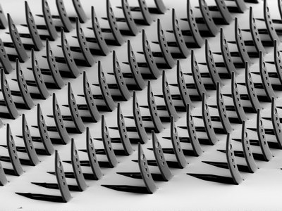
Flexible, high-density microelectrodes in three-dimensional shapes — inspired by the Japanese paper art of kirigami. | Copyrights: Forschungszentrum Jülich
Traditional 3D MEAs tend to rely on rigid materials like silicon, or require intricate manual folding. The new approach, by contrast, is based on flexible, biocompatible materials that minimise the risk of adverse tissue responses. The team uses a polymer film just a few micrometres thick — as supple as cling film, yet robust enough to function as a brain probe. Once placed between a customised mould pair, the flat structure is transformed into its final 3D shape using heat and pressure.
“What continues to surprise me,” says Viviana Rincón Montes, corresponding author and scientist at IBI-3, “is how well a technique designed for macroscopic shaping — like compression moulding — can be scaled down to work so precisely at the microscopic level. Our structures are resilient enough to endure both the mechanical stress of implantation and the biological environment they enter.”
From lab bench to living brain
The team first put their probes through rigorous lab tests to assess their electrochemical performance, folding accuracy and mechanical durability. They were then trialled on brain slices from epilepsy patients and in live mice. The probes successfully captured signals both at the surface and deeper in the brain — including epileptiform activity in human tissue and sensory responses to touch and light in the animal model.
With its high spatial resolution, single-step implantation process and flexibility, the technology is seen as particularly promising for brain–computer interfaces and future therapeutic applications.
Looking ahead: medical applications on the horizon
In the longer term, this technology could help pave the way for visual prosthetics and other neurotechnological innovations. The large number of integrated electrodes not only allows for detailed brain signal recording, but could also make targeted stimulation possible — for example in the retina or visual cortex.
“We’re now working to further optimise the electrode coating and to miniaturise the circuitry,” says Rincón Montes. “Our aim is to create an implant that’s as small, light and efficient as possible — one that can reliably read all 512 electrodes or more.”
While there’s still a long road ahead before clinical use becomes reality, this work lays a solid foundation — combining cutting-edge technology with precision craftsmanship at a microscopic scale.
Kirigami is a variation of origami, the traditional Japanese art of paper folding. Unlike origami, kirigami involves cutting the paper as well — enabling intricate, three-dimensional designs to emerge from the flat surface, all without glue.