Can a person’s individual traits be determined from their brain? For a long time, this was unclear, partly because there were no suitable methods of investigation. Artificial intelligence and big data are now opening up new possibilities and have heralded a change of perspective in brain research.
A human brain is about the size of a head of cauliflower. Inside our brains, approximately 86 billion neurons are connected to each other via 100 trillion interfaces. These are impressive figures, but they do not reflect the whole truth – because they are only averages.
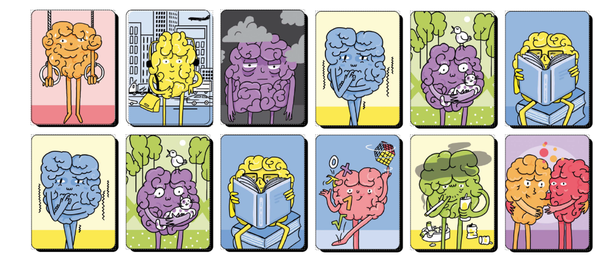
Every person has an individual brain that makes them unique, just as people differ in other characteristics and traits: some have large ears, others small ones; one person may be a thickset, emotional type, another a wiry sports enthusiast. Like every person, each brain is one of a kind. Brain research is only now learning to describe and understand the differences in the human brain properly.
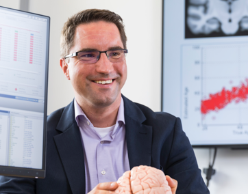
“A shift in perspective is currently taking place. We are no longer focusing on a hypothetical average brain; today, we are much more interested in the differences between individual brains,” says Prof. Simon Eickhoff from the Institute of Neuroscience and Medicine (INM-7). His research focuses on using the brain structure of individual people to draw conclusions about their personal characteristics, as well as diseases such as Alzheimer’s, schizophrenia, or ADHD, and about psychological parameters – such as working memory, spatial awareness, and personality traits.”
For around 150 years, researchers have been trying to link a person’s characteristics to the anatomy of their brain. But this has only recently become possible – thanks to big data methods and machine learning. The data for this are provided by modern imaging techniques, especially magnetic resonance imaging (MRI). This method delivers detailed, three-dimensional images from inside the head. And with functional MRI, it is even possible to visualize which regions of the brain are currently active. This allows researchers to quite literally watch the brain at work.
In order to make reasonable statements about individual cases, the algorithm must first ‘take a look at’ a large number of brains.
Individual data matrix
“We use brain scans to determine the relative sizes or connectivity profiles of several hundred brain regions. These values differ from person to person. This gives us an individual data matrix for each test subject,” explains Eickhoff. Additional information may include, for example, diagnoses, the severity and duration of a disease, or measures of mental performance.
“In the past, imaging was complex, expensive, and not yet widely used. It was therefore only possible to rely on small groups of test subjects – 10 to 20 people who suffered from Parkinson’s disease, for example. Researchers then looked for differences in the brain between the patient group and a healthy control group,” says the neuroscientist. However, the number of test subjects was usually too small to detect typical changes in the patients’ brains.
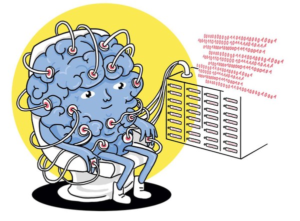
But now, thanks to the wider use of MRI technology, Eickhoff’s team has access to cohort studies, some of which involved many thousands of test subjects. There are hundreds of data points for each patient. Artificial intelligence and big data make it possible to analyse these enormous amounts of information. Eickhoff: “Over the past ten to fifteen years, we have experienced an explosion in the field of machine learning. This has also enabled brain research to take a giant leap forward.”
AI algorithms help the team search these extensive data sets for relevant patterns – complex patterns that would have otherwise remained hidden to the human eye or even classical statistical methods. With each additional training data set, these patterns become increasingly clear. And from this, predictions can be derived for individual patients.
It is not only neurodegenerative conditions such as Alzheimer’s and Parkinson’s that can be diagnosed in this way from brain scans. Similar approaches are also possible for autism spectrum disorder or ADHD. Someone being treated with antidepressants for depression could receive an assessment from the intelligent algorithms on whether it is safe to discontinue the medication – or whether there is a risk of relapse.
“The aim of our work is to use the trained model to make statements about patients whose data were not included in the training. We can diagnose a disease such as Parkinson’s based on a brain scan. And perhaps even make a prognosis about the future course of the disease,” says Eickhoff.
Another piece of the puzzle
It is important to note that this is an assessment based on the patterns observed in the imaging, not a 100 % accurate statement. However, Eickhoff argues that such absolute certainty is not necessarily required for psychiatric practice: “AI provides the doctor with additional information, but it does not make the decision alone. Its judgement is just one piece of the puzzle for the diagnosis.”
How reliable the model is in making the right assessment depends primarily on the quantity and quality of the training data. “In order to make reasonable statements about individual cases, the algorithm must first ‘take a look at’ a large number of brains. It thus learns to filter out, from the many small differences, the features that are truly important for predictions,” says Eickhoff.
The training data not only need to cover a wide range of brain structures, but also, for example, take ethnic factors into account. If they do not, there is a risk of unintended discrimination against minorities. The team at INM-7 tested this with US brain scans: first they trained the AI algorithm exclusively with data from a single, large population group. Then they had the AI analyse data from a minority group. It quickly became apparent that the AI model produced various biases and errors. Eickhoff: “If training data only cover a portion of the population, there is a risk that the model will not recognize all relevant factors. You always have to expect that influencing factors will vary between different population groups, and in some cases significantly.”
Factoring in life circumstances
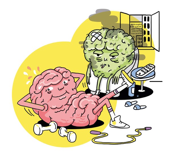
A particular challenge for the researchers is external influencing factors – namely, individual life circumstances: What is the socio-economic background of the test subjects? What kind of environment do they live in? What access to education do they have? What social contacts do they maintain? Do they exercise? Do they read a lot? Have they experienced trauma?
“All of these factors are interconnected and have an influence on the development of our brain – and therefore the things we want to predict: predispositions to disease, cognitive abilities, and personality traits. If this is not adequately taken into account, it can distort the AI model and thus the results,” says Eickhoff.
One way forward could be to further refine the AI models by feeding them not only with the actual training data, but also with preliminary information about the test subjects, such as their gender or socio-cultural background. “This is currently an important research question for us, in which we will need to invest a great deal of work,” says Eickhoff.
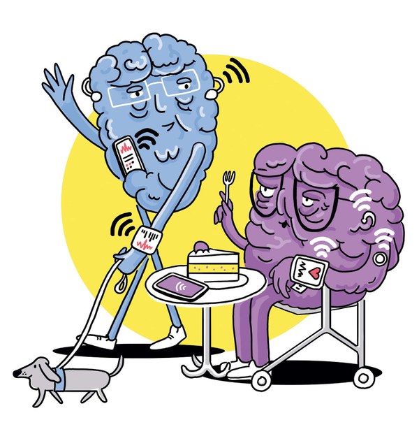
Mobile health and brain research: MRI brain scans provide valuable information for the diagnosis of neurodegenerative diseases such as Alzheimer’s or Parkinson’s (see “Knowing-it-all”). But such scans are complex and expensive – and they only provide a snapshot in time. Wearables such as smartphones, smartwatches, and fitness trackers may not provide brain scans, but they could enable near-continuous monitoring of patients in their everyday lives. One example is the Jülich JTrack apps developed as part of the ABCD-J project for mobile health in North Rhine-Westphalia. The apps record detailed data on patients’ behaviour and impairments in the form of questionnaires or sensor data. For example, the smartphone’s acceleration sensor can measure the tremor of an outstretched arm – a potential early sign of Parkinson’s disease. Artificial intelligence can be used to evaluate such information and thus detect neurological and psychiatric diseases at an early stage.
This text is taken from the 2/25 issue of effzett. Text: Artur Denning, Illustrations: SeitenPlan / Diana Köhne, Image: Forschungszentrum Jülich