

Start-up to be: Clean H2eat
The PET Pioneer
Pioneers – Lifetime achievement of Prof. Langen
Professor Karl-Josef Langen has advanced brain tumor diagnostics. His work is a blueprint for how basic research can evolve into medical innovation. In this interview, he reflects on key successes, obstacles, and current challenges.
May 2025
Imaging technologies are well-established tools in modern medicine, allowing physicians to visualize internal structures of the body—ultrasound and X-rays being familiar examples. Professor Karl-Josef Langen specializes in a more specific field: nuclear medicine imaging, particularly in the diagnosis of brain tumors. These methods involve introducing mildly radioactive substances—so-called tracers—into the body.
Since the mid-1980s, Langen and his team have developed groundbreaking techniques in close collaboration with clinics—methods that today benefit thousands of patients. After decades at Forschungszentrum Jülich, Langen is now retiring. However, he remains active in science, continuing to contribute his expertise at the University Hospital RWTH Aachen.
Prof. Langen, what led you to medical imaging?
During my medical studies, I took a radiology elective and was introduced to imaging technologies like CT and ultrasound. These were still relatively new in the 1980s but held great promise. When I joined Forschungszentrum Jülich in 1985, I began specializing in nuclear medicine.
The pivotal moment came in 1988, at a symposium where I first learned about new applications of positron emission tomography—PET for short. They were using radiolabeled amino acids to diagnose brain tumors. I immediately recognized the enormous potential of this approach.

»To date, we have examined over 11,000 patients, with around 700 more being examined every year. «
Prof. Karl-Josef Langen
Group leader at the Institute of Neuroscience and Medicine: Physics of Medical Imaging (INM-4)
What makes PET imaging with these tracers so effective?
Using radioactive tracers allows us to visualize metabolic activity in the brain. Since cancerous tissue exhibits different metabolic patterns than healthy tissue, PET imaging provides crucial clues about the presence and nature of tumors.
Effzett: Understanding the PET Scan Method
Looking back, which achievement makes you especially proud?
One major milestone was the development of the tracer FET (fluoroethyl-tyrosine) in the mid-1990s at Jülich. FET had key advantages over earlier tracers—particularly in terms of stability.
In 2005, we demonstrated that FET PET could more accurately depict the extent of brain tumors compared to magnetic resonance imaging (MRI). FET PET is now used worldwide and helps distinguish between scar tissue and recurrent tumor growth after treatment—a crucial advantage for patient care.
Detecting brain tumors quickly and precisely: PET and AI
How important has clinical collaboration been to your research?
Absolutely essential. New diagnostic methods only gain acceptance if physicians see clear benefits for their patients. We’ve examined over 11,000 patients so far, and nearly 700 more come to us every year. At the University Hospital RWTH Aachen, we’re a referral center for nearly a third of Germany’s brain tumor cases. Through direct patient contact, we not only witness the real-world impact of our research, but we also receive valuable insights that help us improve our methods.
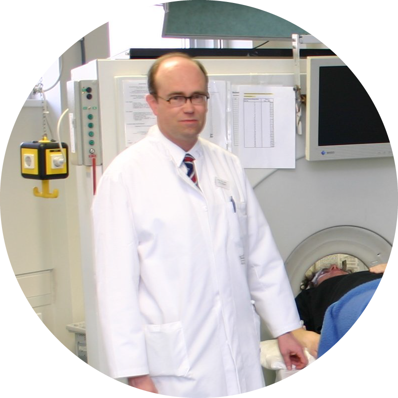
Throwback to the year 2005
Prof. Langen at the PET-Scanner
What are the biggest obstacles in bringing research findings into clinical practice?
The biggest challenges are regulatory requirements and bureaucratic processes. Clinical trials for new radiopharmaceuticals often require many years of research, significantly delaying implementation. PET imaging with new tracers can also be costly, placing financial pressure on many hospitals. Thanks to the infrastructure in Jülich, we were able to offer these examinations for free during the early phases, which helped build acceptance.
What are the current research frontiers in medical imaging?
Artificial intelligence is opening up entirely new possibilities. AI can help analyze PET and MRI images more effectively and automate complex evaluation processes. Our group is already deeply involved in this area. Additionally, the development and evaluation of new tracers remains an exciting and highly relevant research field.
Learn more about nuclear medicine brain tumor diagnostics at the FZ Jülich

Greater Insight, Smarter Decisions
FET PET is a nuclear medicine imaging procedure that visualizes the amino acid metabolism in the brain. It can often depict brain tumors more accurately than a conventional MRI and also helps to detect metastases that have spread to the brain from other parts of the body. In this way, FET PET helps to make more targeted diagnoses and better plan treatments.
Image: Karl-Josef Langen/ Forschungszentrum Jülich

Dive deeper into the current issue
When research, industry, and society unite their perspectives, they create solutions greater than the sum of their parts.
In the Endeavours magazine, discover how co-creation works — through real stories of collaboration, pioneering spirit, and successful transfer.
More articles


Service: Transfer at FZ Jülich


Last Modified: 15.07.2025